

Review
doi: 10.1101/cshperspect.a002477.
Forming and interpreting gradients in the early Xenopus embryo
Affiliations
- PMID: 20066079
- PMCID: PMC2742078
- DOI: 10.1101/cshperspect.a002477
Item in Clipboard
Review
Forming and interpreting gradients in the early Xenopus embryo
Cold Spring Harb Perspect Biol.
2009 Jul.
Abstract
The amphibian embryo provides a powerful model system to study morphogen gradients because of the ease with which it is possible to manipulate the early embryo. In particular, it is possible to introduce exogenous sources of morphogen, to follow the progression of the signal, to monitor the cellular response to induction, and to up- or down-regulate molecules that are involved in all aspects of long-range signaling. In this article, I discuss the evidence that gradients exist in the early amphibian embryo, the way in which morphogens might traverse a field of cells, and the way in which different concentrations of morphogens might be interpreted to activate the expression of different genes.
Figures
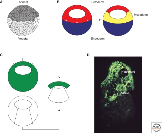
Mesoderm induction. (A) Drawing of a Xenopus embryo at the midblastula stage (Nieuwkoop 1956). The animal hemisphere is to the top and the vegetal hemisphere to the bottom. The mesoderm forms in the equatorial region of the embryo between the two. (B) Mesoderm induction. A signal from the vegetal hemisphere of the embryo (white arrows) causes equatorial cells to form mesoderm. (C) Demonstration of mesoderm induction. Animal pole tissue derived from an embryo uniformly labeled with a fluorescent lineage label is juxtaposed with vegetal pole tissue from an unlabeled embryo and the resulting conjugate is cultured for 3 days. (D) The result of such an experiment. The fluorescently labeled tissue differentiates as notochord and muscle rather than epidermis. Photograph courtesy of Les Dale and Jonathon Slack.
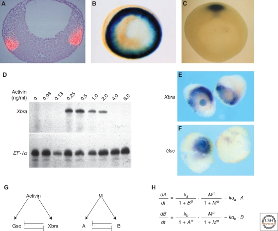
Threshold formation. (A,B) Normal expression of Xbra. (A) Sectioned in situ hybridization showing expression of Xbra in red. Animal pole is to the top and vegetal pole to the bottom. Photograph courtesy of Linda Essex and Michael Sargent. (B) Whole-mount in situ hybridization viewed from the vegetal pole. Notice expression of Xbra throughout the marginal zone. (C) Whole-mount in situ hybridization showing expression of goosecoid in the dorsal region of the embryo. Photograph by Susie Zoltewitz. (http://www.xenbase.org/xenbase/original/WWW/Marker_pages/organizer/goosecoid.html ). (D) Differential expression of Xbra in response to different concentrations of activin. Disaggregated animal pole cells were exposed to the indicated concentrations of activin, reaggregated, and cultured until the equivalent of early gastrula stage 10. They were then assayed for expression of Xbra and EF-1α by RNase protection. Note that Xbra is only activated by intermediate concentrations of activin. (E) Activation of Xbra by intermediate concentrations of activin. An activin-soaked bead was placed between two animal pole regions and cultured for 5 hours before being assayed for expression of Xbra. Note that Xbra is not activated close to the bead but at a distance, where the concentration of activin is at the appropriate level (Papin and Smith 2000). (F) Activation of goosecoid by high levels of activin. An identical experiment to that illustrated in (E), but assaying for expression of goosecoid (Papin and Smith 2000). (G,H) An attempt to understand threshold formation. (G) Left-hand side illustrates that activin activates both goosecoid and Xbra, and the two gene products repress each other's expression. The same relationshiop with activin represented as M (morphogen) and goosecoid and Xbra as “A” and “B”, respectively. (H) Equations describing the rates of synthesis of A and B in response to M. M, A, and B represent the concentrations of each component, ka and kb are the synthesis rates of A and B, respectively, kda and kdb are the decay rates, α and μ are the cooperativities of repression of A by B and B by A, respectively, and μ is the cooperativity of induction by M (Saka and Smith 2007).
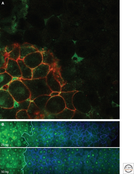
Following long-range signaling in the Xenopus embryo. (A) Animal pole tissue expressing enhanced green fluorescent protein (EGFP)-tagged Xnr2 (cells with red membranes) was juxtaposed with unlabeled tissue and cultured for 2.5 hours. Note the presence of EGFP fluorescence (green) in the unlabeled tissue. Photograph courtesy of Hugh Williams. (B) Visualising activation of the activin signal transduction pathway by Smad2/4 bimolecular fluorescence complementation (BiFC). Animal pole regions derived from embryos previously injected with 25 ng (top) or 50 ng (bottom) mRNA encoding activin (left-hand side) were juxtaposed with tissue derived from embryos expressing GPI-CFP as a membrane-bound lineage marker (right-hand side). Both tissues were also expressing Smad2/4 BiFC constructs. Note nuclear fluorescence in cells at a distance from the source of activin, and that the range of signaling is greater when cells are expressing greater levels of activin. Photograph courtesy of Anja Hagemann.
Similar articles
-
Morphogen gradients, positional information, and Xenopus: interplay of theory and experiment.Dev Dyn. 2002 Dec;225(4):392-408. doi: 10.1002/dvdy.10170. Dev Dyn. 2002. PMID: 12454918 Review.
-
Visualizing long-range movement of the morphogen Xnr2 in the Xenopus embryo.Curr Biol. 2004 Nov 9;14(21):1916-23. doi: 10.1016/j.cub.2004.10.020. Curr Biol. 2004. PMID: 15530392
-
Negative regulation of Smad2 by PIASy is required for proper Xenopus mesoderm formation.Development. 2004 Nov;131(22):5613-26. doi: 10.1242/dev.01449. Epub 2004 Oct 20. Development. 2004. PMID: 15496439
-
Temporal dynamics of patterning by morphogen gradients.Curr Opin Genet Dev. 2009 Aug;19(4):315-22. doi: 10.1016/j.gde.2009.05.004. Epub 2009 Jul 10. Curr Opin Genet Dev. 2009. PMID: 19596567 Review.
-
Expression cloning of Xenopus Os4, an evolutionarily conserved gene, which induces mesoderm and dorsal axis.Dev Biol. 2001 Nov 1;239(1):118-31. doi: 10.1006/dbio.2001.0420. Dev Biol. 2001. PMID: 11784023
Cited by
-
Temporal control of BMP signalling determines neuronal subtype identity in the dorsal neural tube.Development. 2013 Apr;140(7):1467-74. doi: 10.1242/dev.090118. Epub 2013 Mar 5. Development. 2013. PMID: 23462473 Free PMC article.
-
Exosomes as secondary inductive signals involved in kidney organogenesis.J Extracell Vesicles. 2018 Jan 23;7(1):1422675. doi: 10.1080/20013078.2017.1422675. eCollection 2018. J Extracell Vesicles. 2018. PMID: 29410779 Free PMC article.
-
Centrin-2 (Cetn2) mediated regulation of FGF/FGFR gene expression in Xenopus.Sci Rep. 2015 May 27;5:10283. doi: 10.1038/srep10283. Sci Rep. 2015. PMID: 26014913 Free PMC article.
-
Quantitative analyses reveal extracellular dynamics of Wnt ligands in Xenopus embryos.Elife. 2021 Apr 27;10:e55108. doi: 10.7554/eLife.55108. Elife. 2021. PMID: 33904408 Free PMC article.
-
WNT signaling memory is required for ACTIVIN to function as a morphogen in human gastruloids.Elife. 2018 Oct 12;7:e38279. doi: 10.7554/eLife.38279. Elife. 2018. PMID: 30311909 Free PMC article.
References
-
- Albano RM, Godsave SF, Huylebroeck D, Van Nimmen K, Isaacs HV, Slack JMW, Smith JC 1990. A mesoderm-inducing factor produced by WEHI-3 murine myelomonocytic leukaemia cells is activin A. Development 110:435–443 - PubMed
-
- Asashima M, Nakano H, Shimada K, Kinoshita K, Ishii K, Shibai H, Ueno N 1990. Mesodermal induction in early amphibian embryos by activin A (erythroid differentiation factor). Wilhelm Roux' Archiv 198:330–335 - PubMed
-
- Birsoy B, Kofron M, Schaible K, Wylie C, Heasman J 2006. Vg 1 is an essential signaling molecule in Xenopus development. Development 133:15–20 - PubMed
-
- Boterenbrood EC, Nieuwkoop PD 1973. The Formation of the Mesoderm in Urodelean Amphibians. V. Its Regional Induction by the Endoderm. Wilhelm Roux' Archiv 173:319–332 - PubMed
Publication types
MeSH terms
Substances
Grants and funding
LinkOut - more resources
Full Text Sources