Review
. 2009 Jul;1(1):a002576.
doi: 10.1101/cshperspect.a002576.

Gap junctions

Affiliations
Review

Gap junctions

Daniel A Goodenough et al. Cold Spring Harb Perspect Biol. 2009 Jul.

Abstract

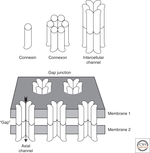
Gap junctions are aggregates of intercellular channels that permit direct cell-cell transfer of ions and small molecules. Initially described as low-resistance ion pathways joining excitable cells (nerve and muscle), gap junctions are found joining virtually all cells in solid tissues. Their long evolutionary history has permitted adaptation of gap-junctional intercellular communication to a variety of functions, with multiple regulatory mechanisms. Gap-junctional channels are composed of hexamers of medium-sized families of integral proteins: connexins in chordates and innexins in precordates. The functions of gap junctions have been explored by studying mutations in flies, worms, and humans, and targeted gene disruption in mice. These studies have revealed a wide diversity of function in tissue and organ biology.


Figures

Figure 1.
Figure 1.
A diagram showing the multiple levels of gap junction structure. Individual connexins assemble intracellularly into hexamers, called connexons, which then traffic to the cell surface. There, they dock with connexons in an adjacent cell, assembling an axial channel spanning two plasma membranes and a narrow extracellular “gap.”
Figure 2.
Figure 2.
Electron microscopy of gap junctions joining adjacent hepatocytes in the mouse. The gap junction (GJ) is seen as an area of close plasma membrane apposition, clearly distinct from the tight junction (TJ) joining these cells. (Inset A) A high magnification view of the gap junction revealing the 2–3 nm “gap” (white arrows) separating the plasma membranes. (Inset B) A freeze-fracture replica of a gap junction showing the characteristic particles on the protoplasmic (P) fracture face and pits on the ectoplasmic (E) fracture face. The particles and pits show considerable disorder in their packing with an average 9-nm center-to-center spacing.
Figure 3.
Figure 3.
A comparison of axial sections through gap-junction structures deduced from X-ray diffraction. The 1985 data (Makowski 1985) were acquired from gap junctions isolated biochemically from mouse liver containing mixtures of Cx32 and Cx26. The intercellular channel (CHANNEL) is blocked at the two cytoplasmic surfaces by electron density at the channel mouths along the sixfold symmetry axis. The 2009 data (Maeda et al. 2009), acquired from three-dimensional crystals of recombinant Cx26, resolve this density at the channel opening as the amino-termini of the connexin proteins, the 2009 model possibly showing an open channel structure.

References

    1. Abrams CK, Freidin MM, Verselis VK, Bargiello TA, Kelsell DP, Richard G, Bennett MV, Bukauskas FF 2006. Properties of human connexin 31, which is implicated in hereditary dermatological disease and deafness. Proc Natl Acad Sci 103:5213–5218. - PMC - PubMed
    1. Albertini DF, Anderson E 1975. Structural modifications of lutein cell gap junctions during pregnancy in the rat and the mouse. Anat Rec 181:171–194 - PubMed
    1. Alexopoulos H, Bottger A, Fischer S, Levin A, Wolf A, Fujisawa T, Hayakawa S, Gojobori T, Davies JA, David CN, et al. 2004. Evolution of gap junctions: The missing link? Curr Biol 14:R879–R880 - PubMed
    1. Allen MJ, Godenschwege TA, Tanouye MA, Phelan P 2006. Making an escape: Development and function of the Drosophila giant fibre system. Semin Cell Dev Biol 17:31–41 - PubMed
    1. Altevogt BM, Paul DL 2004. Four classes of intercellular channels between glial cells in the CNS. J Neurosci 24:4313–4323 - PMC - PubMed

Publication types

LinkOut - more resources