Review
. 2009 Jul;1(1):a002873.
doi: 10.1101/cshperspect.a002873.

Modular design of immunological synapses and kinapses

Affiliations
Review

Modular design of immunological synapses and kinapses

Michael L Dustin. Cold Spring Harb Perspect Biol. 2009 Jul.

Abstract

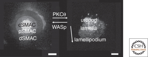
The concept of an immunological synapse goes back to the early 1980s with the discovery of the relationship between T-cell antigen receptor mediated Ca(2+) signaling, adhesion, and directed secretion. However, this concept did not gain traction until images were published starting in 1998 that revealed a specific molecular pattern in the interface between T cells and model antigen-presenting cells or supported planar bilayers. The dominant pattern, a ring of adhesion molecules surrounding a central cluster of antigen receptors, was observed in both model systems. Analysis of the origins of this pattern over the past 10 years has presented a solution for a difficult problem in lymphocyte biology--how a highly motile cell can suddenly stop when it encounters a signal delivered by just a few antigenic ligands on the surface of another cell without disabling the sensory machinery of the motile cell. The T lymphocyte actively assembles the immunological synapse pattern following a modular design with roots in actin-myosin-based motility.


Figures

Figure 1.
Figure 1.
Cytotoxic T lymphocyte (CTL) life cycle. Naïve cells hunt for evidence of infection on the surface of self dendritic cells in lymphoid tissue. One antigen, if found on the primary immunological synapse, leads to activation, proliferation, and development of a CTL. This CTL exits the lymph node and uses the blood to reach a tissue. In the tissue, the CTL migrates to find its target and then uses a secondary synapse to kill the target.
Figure 2.
Figure 2.
SMAC and kinapse zones. Immunological synapses may have a central cSMAC, an annular pSMAC, and an outer dSMAC. The cSMAC can be rich in TCR–peptide–MHC interaction. The pSMAC is enriched in LFA-1–ICAM-1 interactions. These structures are normally in the interface between two cells, but can be modeled using a live T cell interacting with a supported planar bilayer. A kinapse is a mobile junction with the arrow reflecting the direction of motion. The leading edge is the lamellipodium, the wide region is the lamella or focal zone, and the trailing edge is the uropod. These zones are shaded over images showing the pattern of T-cell receptors.
Figure 3.
Figure 3.
Supported planar bilayers. Supported planar bilayers have been used to model the immunological synapse. A remarkable property of planar bilayers is that the lipids, and anything linked to them, are laterally mobile. The left image shows single CD58 molecules in a bilayer labeled with 100 nm fluorescent latex spheres. The left image is based on running background subtraction with peak detection from a 30-s movie and illustrates the areas covered by single diffusing particles in the bilayer. Each particle covers roughly the area of an immunological synapse in 30 s.
Figure 4.
Figure 4.
Reconstitution of the immunological synapse with a planar bilayer. The image on the far right is a slice from the interface between B cells and T cells showing the cSMAC and pSMAC. The schematic and image on the left are of the planar bilayer containing only ICAM-1 and peptide-MHC. The same pattern is formed, although the contact synapse area is larger on the planar surface. The dSMAC was not labeled in these images.
Figure 5.
Figure 5.
Snap shots of TCR microclusters in an immunological synapse. The images are frames from a movie showing the colocalization of TCR and the kinase ZAP-70 (tagged with GFP) as detected by Campi and Varma by TIRFM microscopy (illustrated schematically on the left).

References

    1. Al-Alwan MM, Liwski RS, Haeryfar SM, Baldridge WH, Hoskin DW, Rowden G, West KA 2003. Cutting edge: Dendritic cell actin cytoskeletal polarization during immunological synapse formation is highly antigen-dependent. J Immunol 171:4479–4483 - PubMed
    1. Allen CD, Okada T, Tang HL, Cyster JG 2007. Imaging of germinal center selection events during affinity maturation. Science 315:528–531 - PubMed
    1. Allenspach EJ, Cullinan P, Tong J, Tang Q, Tesciuba AG, Cannon JL, Takahashi SM, Morgan R, Burkhardt JK, Sperling AI 2001. ERM-dependent movement of CD43 defines a novel protein complex distal to the immunological synapse. Immunity 15:739–750 - PubMed
    1. Alon R, Dustin ML 2007. Force as a facilitator of integrin conformational changes during leukocyte arrest on blood vessels and antigen-presenting cells. Immunity 26:17–27 - PubMed
    1. Bachmann MF, McKall-Faienza K, Schmits R, Bouchard D, Beach J, Speiser DE, Mak TW, Ohashi PS 1997. Distinct roles for LFA-1 and CD28 during activation of naive T cells: Adhesion versus costimulation. Immunity 7:549–557 - PubMed

LinkOut - more resources