Review
. 2009 Aug;1(2):a001891.
doi: 10.1101/cshperspect.a001891.

Symmetry breaking during Drosophila oogenesis

Affiliations
Review

Symmetry breaking during Drosophila oogenesis

Siegfried Roth et al. Cold Spring Harb Perspect Biol. 2009 Aug.

Abstract

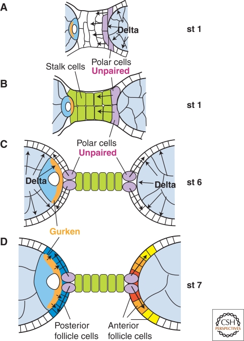
The orthogonal axes of Drosophila are established during oogenesis through a hierarchical series of symmetry-breaking steps, most of which can be traced back to asymmetries inherent in the architecture of the ovary. Oogenesis begins with the formation of a germline cyst of 16 cells connected by ring canals. Two of these 16 cells have four ring canals, whereas the others have fewer. The first symmetry-breaking step is the selection of one of these two cells to become the oocyte. Subsequently, the germline cyst becomes surrounded by somatic follicle cells to generate individual egg chambers. The second symmetry-breaking step is the posterior positioning of the oocyte within the egg chamber, a process mediated by adhesive interactions with a special group of somatic cells. Posterior oocyte positioning is accompanied by a par gene-dependent repolarization of the microtubule network, which establishes the posterior cortex of the oocyte. The next two steps of symmetry breaking occur during midoogenesis after the volume of the oocyte has increased about 10-fold. First, a signal from the oocyte specifies posterior follicle cells, polarizing a symmetric prepattern present within the follicular epithelium. Second, the posterior follicle cells send a signal back to the oocyte, which leads to a second repolarization of the oocyte microtubule network and the asymmetric migration of the oocyte nucleus. This process again requires the par genes. The repolarization of the microtubule network results in the transport of bicoid and oskar mRNAs, the anterior and posterior determinants, respectively, of the embryonic axis, to opposite poles of the oocyte. The asymmetric positioning of the oocyte nucleus defines a cortical region of the oocyte where gurken mRNA is localized, thus breaking the dorsal-ventral symmetry of the egg and embryo.


Figures

Figure 1.
Figure 1.
Loss of polarity along both main-body axes in gurken mutants. Left: egg shell preparation. Right: schematic drawing of eggs showing the orientation of the main body axes, the localization of bicoid and oskar mRNA and the oocyte nucleus.
Figure 2.
Figure 2.
The ovariole. Top: schematic drawing of ovariole with the germarium at the anterior tip and egg chambers of increasing age. Bottom: Magnified view of germarium and stage 9 egg chamber.
Figure 3.
Figure 3.
Cyst formation and oocyte determination. (A) Second cystocyte division producing a four-cell cyst. At metaphase, one spindle pole attaches to the old fusome. Cystokinesis is incomplete, leaving the cystocytes connected by ring canals. Within the ring canals, new fusome material forms. The ring canals move toward each other, leading to clustering of the cystocytes. (B) Oocyte determination. In region 2a of the germarium, the cystocytes are connected by the branched fusome. Both cells with four ring canals (pro-oocytes) show signs of oocyte specification. Later, the fusome is replaced by a polarized microtubule (MT) network emanating from one microtubule organizing center (MTOC) that resides in one of the two pro-oocytes. It is positioned in the region of the Balbiani body, anterior to the oocyte nucleus.
Figure 4.
Figure 4.
The role of polar and cell stalk for axis formation. The region connecting two egg chambers is schematically depicted with anterior and the younger egg chamber pointing to the left side. (A) In region 3 of the germarium, Delta signals from the germline to specify the anterior polar cells. These in turn express the JAK/STAT signaling ligand Unpaired and signal toward the anterior prestalk cells to induce the stalk cell fate. (B) A two-cell-wide stalk forms. Adhesive interactions between the oocyte of the younger cyst and the stalk cells tightly position the oocyte at the posterior pole. The first round of oocyte repolarization takes place indicated by the shift of mRNAs (orange crescent) from the anterior to the posterior pole of the oocyte. (C) At stage 6, a second round of Delta signaling from the germline induces the differentiation of the epithelial follicle cells, which acquire competency to react to a gradient of Unpaired emanating from the polar cells and to Gurken signaling emanating from the oocyte. (D) Unpaired induces terminal cell fates: In the absence of Gurken signaling, the three types of anterior follicle cells form (red, orange, and yellow); in the presence of Gurken signaling, the posterior follicle cells form (blue).
Figure 5.
Figure 5.
Oocyte nucleus migration and dorsoventral axis formation. Schematic drawings of stage 7 oocytes (left) and mature eggs. Top: Wild type. The posterior-to-anterior movement of the oocyte nucleus forces the nucleus to acquire an asymmetrical position, which determines the dorsal side of the egg and establishes orthogonality between the AP and the DV axes. Middle: If nuclear movement does not occur, AP and DV axes are parallel to each other. Bottom: In binuclear oocytes, both nuclei move to the anterior pole to adopt random position at the anterior cortex. Both nuclei induce dorsal egg shell structures.

Similar articles

Cited by

References

    1. Assa-Kunik E, Torres IL, Schejter ED, Johnston DS, Shilo BZ 2007. Drosophila follicle cells are patterned by multiple levels of Notch signaling and antagonism between the Notch and JAK/STAT pathways. Development 134:1161–1169 - PubMed
    1. Becam IE, Tanentzapf G, Lepesant JA, Brown NH, Huynh JR 2005. Integrin-independent repression of cadherin transcription by talin during axis formation in Drosophila. Nat Cell Biol 7:510–516 - PubMed
    1. Benton R, St Johnston D 2003. Drosophila PAR-1 and 14-3-3 inhibit Bazooka/PAR-3 to establish complementary cortical domains in polarized cells. Cell 115:691–704 - PubMed
    1. Benton R, Palacios IM, St Johnston D 2002. Drosophila 14-3-3/PAR-5 is an essential mediator of PAR-1 function in axis formation. Dev Cell 3:659–671 - PubMed
    1. Bolivar J, Huynh JR, Lopez-Schier H, Gonzalez C, St Johnston D, Gonzalez-Reyes A 2001. Centrosome migration into the Drosophila oocyte is independent of BicD and egl, and of the organisation of the microtubule cytoskeleton. Development 128:1889–1897 - PubMed

Publication types

LinkOut - more resources