Review
. 2009 Aug;1(2):a002014.
doi: 10.1101/cshperspect.a002014.

Establishing and interpreting graded Sonic Hedgehog signaling during vertebrate neural tube patterning: the role of negative feedback

Affiliations
Review

Establishing and interpreting graded Sonic Hedgehog signaling during vertebrate neural tube patterning: the role of negative feedback

Vanessa Ribes et al. Cold Spring Harb Perspect Biol. 2009 Aug.

Abstract

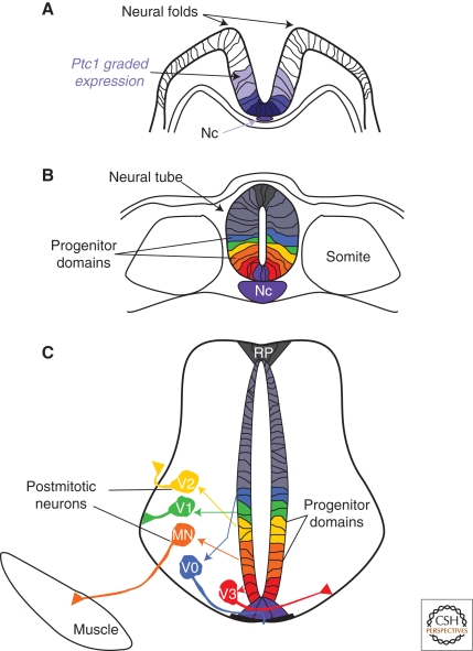
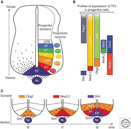
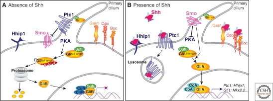
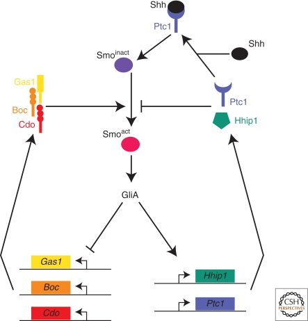
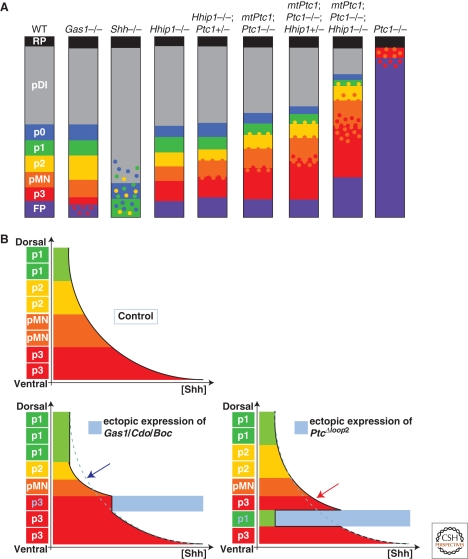
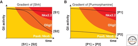
The secreted protein Sonic Hedgehog (SHH) acts in graded fashion to pattern the dorsal-ventral axis of the vertebrate neural tube. This is a dynamic process in which increasing concentrations and durations of exposure to SHH generate neurons with successively more ventral identities. Interactions between the receiving cells and the graded signal underpin the mechanism of SHH action. In particular, negative feedback, involving proteins transcriptionally induced or repressed by SHH signaling, plays an essential role in shaping the graded readout. On one hand, negative feedback controls, in a noncell-autonomous manner, the distribution of SHH across the field of receiving cells. On the other, it acts cell-autonomously to convert different concentrations of SHH into distinct durations of intracellular signal transduction. Together, these mechanisms exemplify a strategy for morphogen interpretation, which we have termed temporal adaptation that relies on the continuous processing and refinement of the cellular response to the graded signal.


Figures

Figure 1.
Figure 1.
Establishment of the spatial organization of neurons from ventral progenitor cells of the spinal cord. (A) Graded expression of the receptor Ptc1 (blue) in the folding neural plate, induced by SHH secreted from the Notochord. (B) After neural tube closure, six domains of progenitor cells, termed FP, p3, pMN, p2, p1, and p0 (purple, red, orange, yellow, green, blue) are precisely arranged along the dorsal–ventral (DV) axis of the neural tube. (C) Each progenitor domain generates a distinct subtype of interneurons (V0–V3) or motor neurons (MN). These postmitotic cells reach their final settling positions within the spinal cord via stereotypic migration pathways. The extension of their axons along specific routes initiates the formation of functional circuits. (Nc) Notochord, (FP) floor plate.
Figure 2.
Figure 2.
Progressive emergence of SHH-regulated gene expression profiles within progenitor cells defines neuronal subtype identities in the ventral spinal cord. (A) Arrayed along the dorsal–ventral (DV) axis of the ventral neural tube are six domains of progenitor cells, FP, p3, pMN, p2, p1, and p0, which generate V0–V3 and MN neuronal subtypes. The spatial organization of the progenitor domains is established by a gradient of SHH protein (purple) secreted from the Nc and FP. (B) The restricted expression profiles of the TFs Nkx2.2, Olig2, Nkx6.1, Nkx6.2, Dbx1, Dbx2, Irx3, Pax6, and Pax7 within progenitors is regulated by graded SHH signaling. Each progenitor domain expresses a unique combination of TFs. (C) The TFs Olig2 and Nkx2.2, as well as SHH itself, distinguish the three most ventral progenitor domains (pMN, p3, and FP, respectively). The expression of each of these markers is initiated at successive developmental time points within the midline of the neural tube and extends to more dorsal positions with the appearance of the next marker at the midline. This series of gene induction events occurs in parallel to the accumulation and extension of the gradient of SHH protein in the ventral neural tube. (Nc) Notochord, (FP) floor plate.
Figure 3.
Figure 3.
SHH signal transduction. (A) In the absence of SHH, the receptor Ptc1 represses the activity of the transmembrane protein Smo and the translocation of Smo to the primary cilium of the cell. In these cells, protein kinase A (PKA) promotes the proteasome-dependent partial processing or complete degradation of the Gli transcription factors (Gli2 and Gli3). The truncated forms of these proteins (GliR) translocate to the nucleus and repress the transcription of SHH signaling targets. SuFu maintains any remaining full-length Gli proteins in an inactive state. (B) The binding of SHH to Ptc1 releases repression of Smo, allowing its translocation into the cell's cilium. The activation of Smo inhibits the proteolytic processing of Gli proteins and culminates in activated Gli proteins (GliA) translocating to the nucleus to activate target gene expression. At the cell surface, in addition to Ptc1, the SHH-binding membrane protein Hhip1 binds SHH and inhibits signaling. Conversely, the SHH-binding proteins Gas1, Cdo, and Boc enhance the response of cells to the morphogen.
Figure 4.
Figure 4.
Negative feedback loops in the SHH signaling pathway driven by SHH-binding proteins. The membrane proteins Gas1, Cdo, and Boc promote the activation of Smo and are transcriptionally inhibited by SHH signaling. Conversely, Ptc1 and Hhip1 are transcriptionally induced by Gli activation and inhibit the transduction of a SHH signal to Smo.
Figure 5.
Figure 5.
Regulation of SHH-mediated pattern formation in the ventral spinal cord by the SHH-binding proteins. (A) Diagrammatic representation of the effect of mutations of SHH binding proteins on pattern formation in the neural tube. SHH−/− spinal cord displays a complete absence of molecular identities characteristic of the FP, p3, and pMN progenitor domains and severe reduction and ventral displacement of p2, p1, and p0 identities. Loss of Gas1 (Gas1−/−) results in a reduction of the number of p3 cells, reminiscent of a mild reduction in the SHH signaling activity. In contrast to Gas1 and SHH mutant embryos, compound mutant embryos for Hhip1 and Ptc1 display expansion of the ventral most progenitor domains. In the Hhip1 mutant, the p3 progenitor domain increases in size. The other ventral progenitor domains remain unaffected. More dramatically, in Ptc1 mutants, dorsal progenitor identities are absent, with the exception of the roof plate, and most progenitor cells adopt a FP identity. A few scattered cells with pMN and p3 characteristics are intermixed in the most dorsal regions. This phenotype is partially rescued by a transgene expressing Ptc1 ubiquitously at low levels (MtPtc1). Thus, in the MtPtc1;Ptc1−/−, the p3, pMN progenitor domains are expanding dorsally. Most strikingly, cells with distinct fates are mixed at the boundaries between the p3 and pMN, and the pMN and the p2 progenitor domains in these compound mutants, indicating a role for Ptc1-mediated feedback in the precision of pattern formation. The progressive loss of alleles of Hhip1 in the MtPtc1;Ptc1−/− background exacerbates these phenotypes, demonstrating that Ptc1 and Hhip1 share redundant functions in defining the position and precision of boundaries between distinct progenitor domains. (B) Schematic representation of the gradient of SHH along the DV axis of the neural tube and of the subsequent position of the ventral cell fates in control embryos or in embryos in which Gas1/Cdo/Boc or Ptc1Δloop2 have been ectopically expressed (blue shading). The expression of Gas1, Cdo, or Boc results in a local increase in the concentration of SHH protein, thus ventralizing the fate of cells. Because of the local sequestration of the ligand, SHH is reduced dorsally (blue arrow). As a consequence, the size of pMN and p1 domains are decreased in size. The expression of Ptc1Δloop2 leads to a cell-autonomous reduction of the response of cells to SHH. A consequence of this is that cells that would normally adopt a p3 identity are transformed to a more dorsal fate. Ptc1Δloop2 does not sequester SHH protein. Therefore, SHH concentration is increased in a noncell-autonomous manner, dorsal to the Ptc1Δloop2 expressing region, which leads to the generation of p3 cell fate at a position where pMN is normally generated.
Figure 6.
Figure 6.
Correlation between the temporal profiles of Gli activity and gene expression profiles in cells exposed to either SHH or the Smo-agonist purmorphamine. (A) Different concentrations of SHH generate distinct durations of Gli activity. Both low (S1) and high (S2) concentrations of morphogen initially induce high levels of Gli activity. Over time, in cells exposed to a low concentration of SHH, the Gli activity drops quickly, whereas at the high concentration of morphogen, Gli activity is maintained at high levels. Consequently, high concentrations of SHH induce Nkx2.2 after transiently expressing Olig2, whereas at the lower concentration, cells maintain Olig2 expression. (B) In contrast to SHH, purmorphamine, a Smo agonist, generates profiles of Gli activity that do not vary as greatly through time. A high concentration (P2) consistently generates higher levels of Gli activity at each time point than a lower concentration (P1). This suggests that the generation of distinct periods of Gli activity by SHH is mediated upstream of Smo.

Similar articles

Cited by

References

    1. Allen BL, Tenzen T, McMahon AP 2007. The Hedgehog- binding proteins Gas1 and Cdo cooperate to positively regulate SHH signaling during mouse development. Genes Dev 21:1244–1257 - PMC - PubMed
    1. Ashe HL, Briscoe J 2006. The interpretation of morphogen gradients. Development 133:385–394 - PubMed
    1. Aza-Blanc P, Lin HY, Ruiz i Altaba A, Kornberg TB 2000. Expression of the vertebrate Gli proteins in Drosophila reveals a distribution of activator and repressor activities. Development 127:4293–4301 - PubMed
    1. Bai CB, Stephen D, Joyner AL 2004. All mouse ventral spinal cord patterning by hedgehog is Gli dependent and involves an activator function of Gli3. Dev Cell 6:103–115 - PubMed
    1. Bailey PJ, Klos JM, Andersson E, Karlen M, Kallstrom M, Ponjavic J, Muhr J, Lenhard B, Sandelin A, Ericson J 2006. A global genomic transcriptional code associated with CNS-expressed genes. Exp Cell Res 312:3108–3119 - PubMed

Publication types

LinkOut - more resources