Review
. 2009 Aug;1(2):a002519.
doi: 10.1101/cshperspect.a002519.

Gradients in the brain: the control of the development of form and function in the cerebral cortex

Affiliations
Review

Gradients in the brain: the control of the development of form and function in the cerebral cortex

Stephen N Sansom et al. Cold Spring Harb Perspect Biol. 2009 Aug.

Abstract

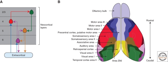
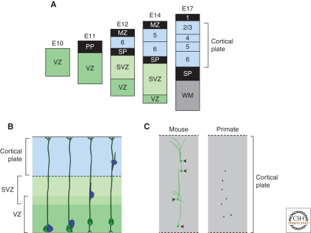
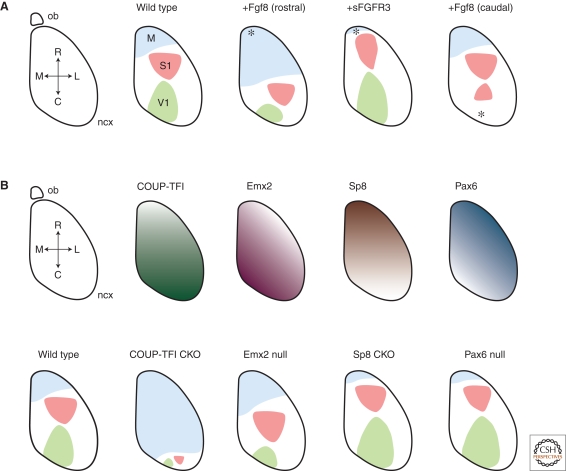
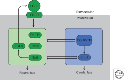
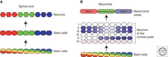
In the developing brain, gradients are commonly used to divide neurogenic regions into distinct functional domains. In this article, we discuss the functions of morphogen and gene expression gradients in the assembly of the nervous system in the context of the development of the cerebral cortex. The cerebral cortex is a mammal-specific region of the forebrain that functions at the top of the neural hierarchy to process and interpret sensory information, plan and organize tasks, and to control motor functions. The mature cerebral cortex is a modular structure, consisting of anatomically and functionally distinct areas. Those areas of neurons are generated from a uniform neuroepithelial sheet by two forms of gradients: graded extracellular signals and a set of transcription factor gradients operating across the field of neocortical stem cells. Fgf signaling from the rostral pole of the cerebral cortex sets up gradients of expression of transcription factors by both activating and repressing gene expression. However, in contrast to the spinal cord and the early Drosophila embryo, these gradients are not subsequently resolved into molecularly distinct domains of gene expression. Instead, graded information in stem cells is translated into discrete, region-specific gene expression in the postmitotic neuronal progeny of the stem cells.


Figures

Figure 1.
Figure 1.
The arrangement of neocortical circuits and areas in the adult mouse brain. (A) The basic cortical circuit. Major extracortical inputs terminate in layer 4 and to a lesser degree in layer 6. Layer 4 neurons project to layers 2 and 3, which in turn innervate layers 5 and 6, the major output layers of the cortex. (B) Dorsal view of the adult mouse brain, with the functional roles of histologically defined areas labeled. Area maps redrawn from Wree et al. 1983.
Figure 2.
Figure 2.
Cortical stem cells are multipotent, generating neurons for each layer in a fixed temporal order. (A) Layer-specific neurons are generated in a fixed temporal order in a classic inside-out pattern over 6 days in the mouse cortex. (B) Neurons (blue) and generated by radial glia stem cells (green) in the ventricular zone and subsequently migrate radially outwards into the cortical plate along the processes of the radial glia cells that span the width of the developing neocortex. (C) Cortical stem cells generate radially arranged clones of neurons in mice and primates. Examples of retrovirally labeled clones are redrawn from Kornack and Rakic 1995 and Yu et al. 2009.
Figure 3.
Figure 3.
The roles of FGF signaling and graded transcription factor expression in neocortical pattern formation. (A) Diagram of a dorsal view of the mouse cortex (ncx, neocortex; ob, olfactory bulb; M, medial; L, lateral; R, rostral; C, caudal), with the major axes and areas labeled (M, motor; S1, somatosensory; V1, primary visual). The effects of altering the levels and positions of FGF8 signaling are shown. Increasing FGF8 levels rostrally increases the size of the rostral motor area at the expense of caudal areas. Conversely, antagonizing FGF8 signaling by expression of the extracellular face of an FGF receptor (sFGFR3) results in a reduction of the size of M1 and an increase in the size of caudal areas. Introduction of a new, caudal source of FGF8 results in the generation of a mirror-image of the S1 area in caudal cortex. (B) The transcription factors COUP-TF1, Emx2, Sp8, and Pax6 are expressed in gradients along the rostrocaudal axis of the cortex as shown. The effects of null mutations in each transcription factor are shown (CKO, cortex-specific knockout). See text for details of each phenotype.
Figure 4.
Figure 4.
The outline of a cellular network for controlling neocortical area formation. FGF8, signaling through FGF receptors, induces or increases expression of the rostrally expressed transcription factors, the ETS factors Pax6 and FGF8. Sp8 in turn increases expression of Fgf8 rostrally. The rostral and caudal transcription factors show a degree of mutual cross-repression. Among the caudal transcription factors, COUP-TFI appears to be upstream of Emx2, as loss of Emx2 does not alter COUP-TFI expression, and COUP-TFI-null cortices have a more severe patterning phenotype than that observed in Emx2 nulls.
Figure 5.
Figure 5.
The translation of a gradient-based neocortical map in stem cells into spatially discrete patterns of neuronal gene expression in the neocortex. (A) In the developing spinal cord, an initial graded pattern of transcription factor expression along the dorsoventral axis is progressively resolved into clearly delineated, molecularly distinct stem cell domains. Each spatially defined group of stem cells goes on to produce different classes of spinal cord neurons. (B) In contrast, in the developing neocortex, gradients of expression in the ventricular zone do not resolve into discrete spatial domains. Instead, those gradients appear to be translated into discrete spatial expression in the neurons generated by those stem cells. Furthermore, the spacial expression of specific genes often differs between different neuronal layers, as shown here for the gene Epha7.

Similar articles

Cited by

References

    1. Anderson SA, Eisenstat DD, Shi L, Rubenstein JL 1997. Interneuron migration from basal forebrain to neocortex: Dependence on Dlx genes. Science 278:474–476 - PubMed
    1. Armentano M, Chou SJ, Tomassy GS, Leingartner A, O'Leary DD, Studer M 2007. COUP-TFI regulates the balance of cortical patterning between frontal/motor and sensory areas. Nat Neurosci 10:1277–1286 - PubMed
    1. Bachler M, Neubüser A 2001. Expression of members of the Fgf family and their receptors during midfacial development. Mech Dev 100:313–316 - PubMed
    1. Barbe MF, Levitt P 1991. The early commitment of fetal neurons to the limbic cortex. J Neurosci 11:519–533 - PMC - PubMed
    1. Bishop KM, Rubenstein JLR, O'Leary DDM 2002. Distinct actions of Emx1, Emx2, and Pax6 in regulating the specification of areas in the developing neocortex. J Neurosci 22:7627–7638 - PMC - PubMed

Substances

LinkOut - more resources