Review
. 2009 Aug;1(2):a002543.
doi: 10.1101/cshperspect.a002543.

The desmosome

Affiliations
Review

The desmosome

Emmanuella Delva et al. Cold Spring Harb Perspect Biol. 2009 Aug.

Abstract

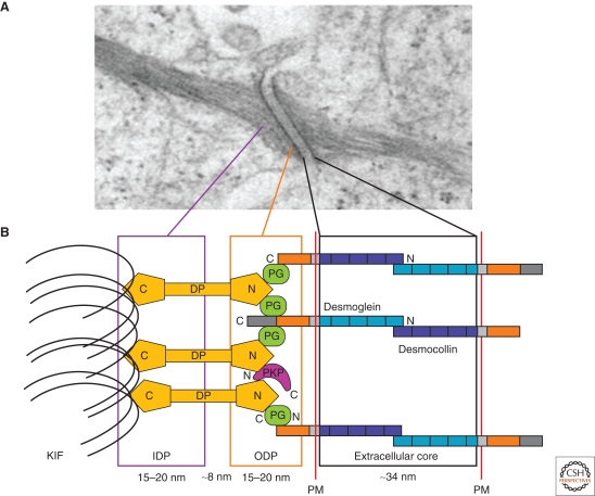
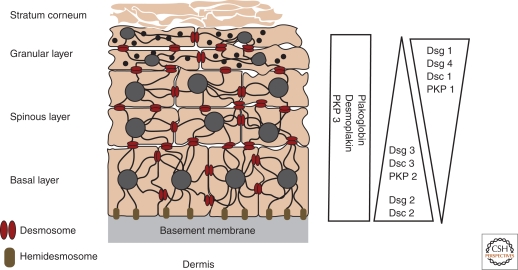
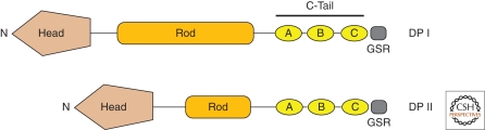
Desmosomes are intercellular junctions that tether intermediate filaments to the plasma membrane. Desmogleins and desmocollins, members of the cadherin superfamily, mediate adhesion at desmosomes. Cytoplasmic components of the desmosome associate with the desmosomal cadherin tails through a series of protein interactions, which serve to recruit intermediate filaments to sites of desmosome assembly. These desmosomal plaque components include plakoglobin and the plakophilins, members of the armadillo gene family. Linkage to the cytoskeleton is mediated by the intermediate filament binding protein, desmoplakin, which associates with both plakoglobin and plakophilins. Although desmosomes are critical for maintaining stable cell-cell adhesion, emerging evidence indicates that they are also dynamic structures that contribute to cellular processes beyond that of cell adhesion. This article outlines the structure and function of the major desmosomal proteins, and explores the contributions of this protein complex to tissue architecture and morphogenesis.


Figures

Figure 1.
Figure 1.
A model for the structure of desmosomes. (A) Electron micrograph of a desmosome. (B) Schematic of desmosomal proteins and relative distance from the plasma membrane (PM). The desmosomal cadherins, the desmogleins and desmocollins, extend into extracellular core and outer dense plaque (ODP) to establish contact and adhere to neighboring cells in a Ca2+-dependent manner. The cadherin cytoplasmic tails associate linker proteins, plakoglobin (PG), the plakophilins (PKP), and desmoplakin (DP). DP binds to keratin intermediate filaments (KIF) within the inner dense plaque (IDP), serving to tether the intermediate filaments to the plasma membrane. (Adapted with permission from Kottke et al. 2006.)
Figure 2.
Figure 2.
Domains of the desmosomal cadherins. Desmogleins (Dsg) and desmocollins (Dsc) contain four highly conserved extracellular (EC) domains, a more variable extracellular anchor (EA), a single transmembrane domain (TM), and an intracellular anchor (IA), which is followed by additional cytoplasmic domains. Desmocollin “a” isoforms and desmogleins contain an intracellular cadherin-like sequence (ICS) that binds plakoglobin. Desmogleins contain the additional intracellular proline-rich linker (IPL), a variable number of repeat unit domains (RUD), and a desmoglein terminal domain (DTD). The numbers of RUDs contained in each desmoglein isoform are as follows: Dsg 1, 5 repeats; Dsg 2, 6 repeats; Dsg 3, 2 repeats; and Dsg 4, 3 repeats.
Figure 3.
Figure 3.
Expression patterns of the desmosomal cadherins in the epidermis. The epidermis is a stratified epithelium comprised of four distinct layers—the basal layer, spinous layer, granular layer, and the stratum corneum. Keratin filaments are shown connecting to desmosomes at sites of cell–cell contact and to hemidesmosomes at the basement membrane. The relative expression profiles of the various desmosomal cadherins and plaque proteins in the epidermal layers are depicted on the right. (Adapted with permission from Kottke et al. 2006.)
Figure 4.
Figure 4.
Armadillo protein family members. Plakoglobin (PG) contains 12-arm repeats that are flanked by distinct amino- and carboxy-termini. The plakophilins (PKP) contain 9-arm repeats with an insert between repeats 5 and 6 that introduces a bend into the overall structure. PKP1 and PKP2 exist as two isoforms, each generating a short “a” form and a longer “b” form, differing by the addition of 21 amino acids in arm repeat three (PKP1) and the addition of 44 amino acids in arm repeat four (PKP2).
Figure 5.
Figure 5.
Desmoplakin. Shown above are the different domains that form the two desmoplakin isoforms, DP I and II. The two isoforms differ in the length of the central rod domain (Rod), with DP II missing approximately two-thirds of the Rod domain. The carboxy-terminal tail (C-Tail) contains three plakin repeat domains (A, B, C), as well as a glycine-serine-arginine rich domain (GSR) thought to regulate desmoplakin binding to intermediate filaments. The amino-terminal globular head domain (Head) mediates protein–protein interactions. See text for additional details.

Similar articles

Cited by

References

    1. Acehan D, Petzold C, Gumper I, Sabatini DD, Muller EJ, Cowin P, Stokes DL 2008. Plakoglobin is required for effective intermediate filament anchorage to desmosomes. J Invest Dermatol 128:2665–2675 - PubMed
    1. Al-Amoudi A, Frangakis AS 2008. Structural studies on desmosomes. Biochem Soc Trans 36:181–187 - PubMed
    1. Amagai M 1996. Pemphigus: Autoimmunity to epidermal cell adhesion molecules. Adv Dermatol 11:319–352; discussion 353 - PubMed
    1. Amagai M 1999. Autoimmunity against desmosomal cadherins in pemphigus. J Dermatol Sci 20:92–102 - PubMed
    1. Amagai M 2002. Pemphigus as a paradigm of autoimmunity and cell adhesion. Keio J Med 51:133–139 - PubMed

Publication types

LinkOut - more resources