Review
. 2009 Oct;1(4):a003087.
doi: 10.1101/cshperspect.a003087.

Listeria monocytogenes internalin and E-cadherin: from bench to bedside

Affiliations
Review

Listeria monocytogenes internalin and E-cadherin: from bench to bedside

Matteo Bonazzi et al. Cold Spring Harb Perspect Biol. 2009 Oct.

Abstract

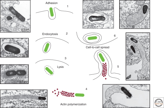
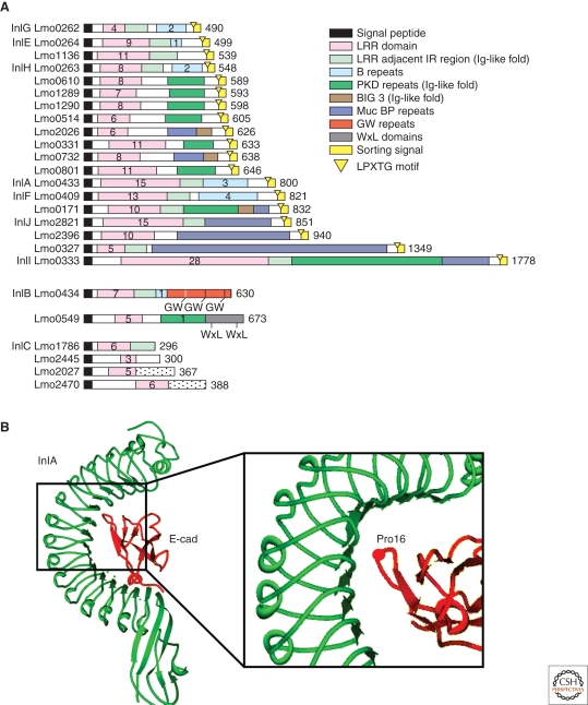
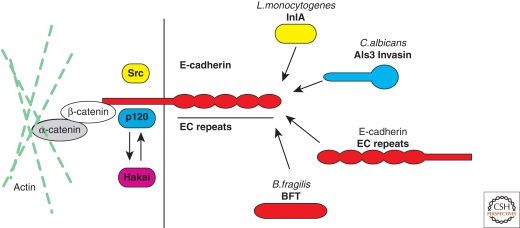
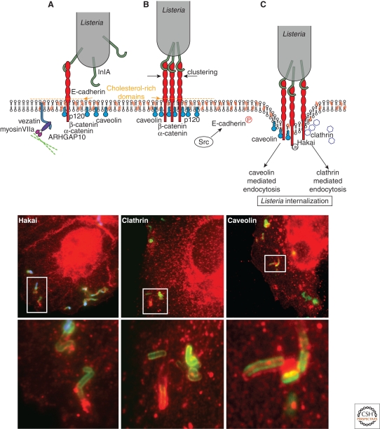
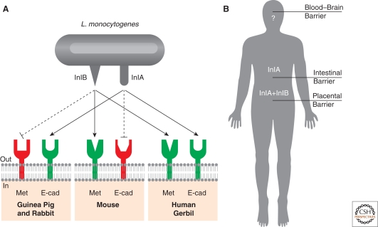
Listeria monocytogenes is a Gram-positive bacterium responsible for a severe infection associated with different clinical features (gastroenteritis, meningoencephalitis, and abortion in pregnant women). These pathologies are caused by the unusual capacity of the bacterium to cross three host barriers during infection and to invade nonphagocytic cells. To invade host cells, Listeria uses two proteins, InlA and InlB, which have specific receptors on the host-cell surface, E-cadherin and Met, respectively. Here, we discuss the specificity of the InlA-E-cadherin interaction, the signaling cascade activated on E-cadherin engagement by InlA, and the role of InlA and E-cadherin in the breaching of host barriers and the dissemination of the infection.


Figures

Figure 1.
Figure 1.
The cell cycle of L. monocytogenes. (1) L. monocytogenes adheres to the surface of epithelial cells via the interactions of the surface proteins InlA and InlB with E-cadherin and the Met receptor, respectively. (2) On internalization, L. monocytogenes is engulfed in a phagocytic vacuole. (3) L. monocytogenes lyses vacuolar membranes by means of the toxin LLO. (4) L. monocytogenes uses the protein ActA to harness the actin polymerization machinery and facilitate its intracellular movement via the formation of so-called actin “comet tails.” (5) L. monocytogenes exploits actin-based motility for direct cell-to-cell spread to allow the dissemination of the infection to neighboring cells via the formation of plasma membrane protrusions. (6) Once internalized by neighboring cells, L. monocytogenes is confined in a double-membrane vacuole from which it escapes to restart its life cycle.
Figure 2.
Figure 2.
The internalin family and InlA. (A) Schematic representation of the internalin family of proteins of L. monocytogenes. Homologous regions are color coded as indicated in the legend. Numbers within different domains indicate the number of repeats (Bierne et al. 2007). (B) Crystal structure of InlA in complex with the EC1 domain of E-cadherin. The LRR domain of InlA consists of fifteen and a half 22-residue repeats that form a right-handed curved solenoid structure. Each repeat begins with a β strand of five residues: these strands combine in a 16-stranded β sheet that forms a pseudo-helical surface. The presence of a proline in position 16 of E-cadherin allows the terminal loop of E-cadherin to be hydrophobic and uncharged, therefore strengthening the interaction with InlA.
Figure 3.
Figure 3.
E-cadherin interactors. E-cadherin is a single-pass transmembrane protein with a 152-amino-acid intracellular domain and a 555-amino-acid extracellular domain. The intracellular domain of E-cadherin interacts with proteins of the catenin family. p120 catenin binds to the juxtamembrane domain of E-cadherin and stabilizes E-cadherin at the plasma membrane. On E-cadherin phosphorylation by the tyrosine kinase Src p120 is released and the ubiquitin-ligase Hakai can bind and ubiquitinate E-cadherin. β-catenin binds to the carboxy-terminal domain of E-cadherin and mediates the interaction with the actin cytoskeleton via α-catenin. The extracellular domain of E-cadherin interacts with the EC domains of E-cadherin molecules from neighboring cells, it serves as the receptor for the L. monocytogenes surface protein InlA and the C. albicans invasin Als3, and is the target of the B. fragilis metalloprotease toxin BFT.
Figure 4.
Figure 4.
Dynamics of the InlA/E-cadherin interaction (upper panel). (A) On engagement of E-cadherin by InlA, the adherens junction machinery is activated, inducing the recruitment of the junctional proteins α-catenin, p120 catenin, ARHGAP10, and myosin VIIa. (B) InlA interaction with E-cadherin induces the caveolin-dependent clustering of E-cadherin and the activation of the tyrosine kinase Src. (C) The Src-dependent phosphorylation of E-cadherin triggers the recruitment of the ubiquitin-ligase Hakai and the ubiquitination of E-cadherin. E-cadherin ubiquitination induces the sorting of E-cadherin within clathrin-coated pits for bacterial internalization. Alternatively, E-cadherin may persist within caveolin-rich domains and bacterial internalization can occur via caveosomes. Bottom panels: Listeria (green) recruits Hakai (left panel), clathrin (middle panel), and caveolin (right panel) at the bacterial entry site when invading epithelial cells. Lower images are a magnified view of the highlighted areas.
Figure 5.
Figure 5.
Species specificity of the InlA-E-cadherin interaction (A) The species specificity of the InlA/E–cadherin interaction depends on the presence of a proline in position 16 of the amino-terminal E-cadherin repeat. Species that substitute a glutamate to proline in position 16 of E-cadherin are not sensitive to Listeria infection, as InlA/E–cadherin interactions cannot occur. Based on the observation that some species are not sensitive to InlB, recent studies revealed that species specificity occurs in the case of InlB. (B) InlA/E-cadherin interaction is fundamental to cross the intestinal barrier, whereas InlB does not play any role at this stage. A coordinated action of the two internalins is necessary to cross the placental barrier, whereas the role of InlA and InlB at the level of the blood–brain barrier is still an unresolved issue.

Similar articles

Cited by

References

    1. Agerer F, Lux S, Michel A, Rohde M, Ohlsen K, Hauck CR 2005. Cellular invasion by Staphylococcus aureus reveals a functional link between focal adhesion kinase and cortactin in integrin-mediated internalisation. J Cell Sci 118:2189–2200 - PubMed
    1. Bakardjiev AI, Stacy BA, Portnoy DA 2005. Growth of Listeria monocytogenes in the guinea pig placenta and role of cell-to-cell spread in fetal infection. J Infect Dis 191:1889–1897 - PubMed
    1. Bierne H, Sabet C, Personnic N, Cossart P 2007. Internalins: A complex family of leucine-rich repeat-containing proteins in Listeria monocytogenes. Microbes Infect 9:1156–1166 - PubMed
    1. Bonazzi M, Cossart P 2006. Bacterial entry into cells: A role for the endocytic machinery. FEBS Lett 580:2962–2967 - PubMed
    1. Bonazzi M, Lecuit M, Cossart P 2009. Listeria monocytogenes internalin and E-cadherin: From structure to pathogenesis. Cell Microbiol 11:693–702 - PubMed

LinkOut - more resources