

Review
doi: 10.1101/cshperspect.a000075.
A structural guide to proteins of the NF-kappaB signaling module
Affiliations
- PMID: 20066103
- PMCID: PMC2773632
- DOI: 10.1101/cshperspect.a000075
Item in Clipboard
Review
A structural guide to proteins of the NF-kappaB signaling module
Cold Spring Harb Perspect Biol.
2009 Sep.
Abstract
The prosurvival transcription factor NF-kappaB specifically binds promoter DNA to activate target gene expression. NF-kappaB is regulated through interactions with IkappaB inhibitor proteins. Active proteolysis of these IkappaB proteins is, in turn, under the control of the IkappaB kinase complex (IKK). Together, these three molecules form the NF-kappaB signaling module. Studies aimed at characterizing the molecular mechanisms of NF-kappaB, IkappaB, and IKK in terms of their three-dimensional structures have lead to a greater understanding of this vital transcription factor system.
Figures

The NF-κB signaling module. NF-κB exists in the cytoplasm of resting cells by virtue of its noncovalent association with an IκB inhibitor protein. The IκB kinase (IKK) responds to diverse stimuli by catalyzing the phosphorylation-dependent 26 S proteasome-mediated degradation of complex-associated IκB. Active NF-κB accumulates in the nucleus where it binds with DNA sequence specificity in the promoter regions of target genes and activates their transcription.
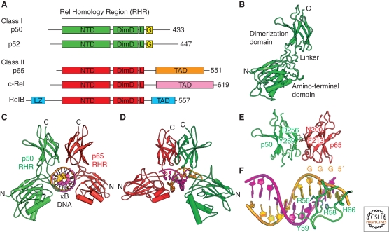
The NF-κB family. (A) The human genome encodes five polypeptides that assemble in various dimer combinations to form active NF-κB transcription factors. Each of the subunits contains the Rel homology region (RHR) near its amino terminus. The RHR consists of two folded domains, the amino-terminal domain (NTD) and the dimerization domain (DimD), that are joined by a short flexible linker and a carboxy-terminal flexible region that contains the nuclear localization signal (L). Three of the subunits, p65, c-Rel, and RelB, also contain a transcription activation domain (TAD) at their carboxy-terminal ends. RelB contains a predicted leucine zipper motif (LZ) amino-terminal to its RHR. The NF-κB subunits p50 and p52 lack transactivation domains and have glycine-rich regions (G). (B) A ribbon diagram representation of the RHR from p50 in its DNA-bound conformation. (C) The NF-κB p50:p65/RelA heterodimer bound to κB DNA. (D) Another view of the complex. (E) The NF-κB p50:p65/RelA heterodimer dimerization domains with key amino acid side chains labeled. (F) κB DNA from the NF-κB:DNA complex with key base-contacting amino acid residues labeled.
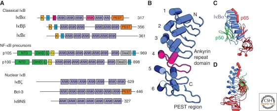
The family of human IκB proteins. (A) IκB proteins are classified as in text. Classical IκB proteins possess ankyrin repeats (ANK) flanked by an amino-terminal signal response region and carboxy terminal PEST region. The signal response regions contain sites of phosphorylation by IKK (S), ubiquitination (K), and nuclear export (E). The NF-κB precursors serve as IκB proteins as well as the source of the mature p50 and p52 NF-κB subunits. (B) Ribbon diagram of the IκBα structure from the NF-κB:IκBα complex crystal structure. Individual ankyrin repeats are numbered, ANK 4 is colored magenta, and the PEST region is labeled. (C) Ribbon diagram of the NF-κB:IκBα complex. (D) Another view of the complex.
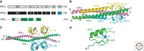
Subunits of the human IKK complex. (A) Domain organization of IKK subunits. Catalytic subunits contain a kinase domain (KD), ubiquitin-like domain (U), leucine zipper (L), helix-loop-helix (H), serine-rich (S), and NEMO-binding motif (N). The NEMO/IKKγ subunit contains two predicted coiled-coil motifs (CC1 and ‐2), a leucine zipper (L), and a carboxy-terminal zinc-finger (ZF). (B) Ribbon diagram of the IKK2/IKKβ:NEMO/IKKγ complex. Individual polypeptides are labeled as well as some of the conserved hydrophobic amino acid side chains from IKK2/IKKβ. (C) Ribbon diagram of the NEMO/IKKγ:di-ubiquitin complex. (D) The NEMO/IKKγ carboxy-terminal zinc-finger motif structure.
Similar articles
-
Regulation and function of IKK and IKK-related kinases.Sci STKE. 2006 Oct 17;2006(357):re13. doi: 10.1126/stke.3572006re13. Sci STKE. 2006. PMID: 17047224 Review.
-
A homeostatic model of IkappaB metabolism to control constitutive NF-kappaB activity.Mol Syst Biol. 2007;3:111. doi: 10.1038/msb4100148. Epub 2007 May 8. Mol Syst Biol. 2007. PMID: 17486138 Free PMC article.
-
IkappaB kinase-independent IkappaBalpha degradation pathway: functional NF-kappaB activity and implications for cancer therapy.Mol Cell Biol. 2003 Nov;23(22):8070-83. doi: 10.1128/MCB.23.22.8070-8083.2003. Mol Cell Biol. 2003. PMID: 14585967 Free PMC article.
-
NF-kappaB p105 is a target of IkappaB kinases and controls signal induction of Bcl-3-p50 complexes.EMBO J. 1999 Sep 1;18(17):4766-78. doi: 10.1093/emboj/18.17.4766. EMBO J. 1999. PMID: 10469655 Free PMC article.
-
Phosphorylation meets ubiquitination: the control of NF-[kappa]B activity.Annu Rev Immunol. 2000;18:621-63. doi: 10.1146/annurev.immunol.18.1.621. Annu Rev Immunol. 2000. PMID: 10837071 Review.
Cited by
-
A New Paradigm to Mitigate Osteosarcoma by Regulation of MicroRNAs and Suppression of the NF-κB Signaling Cascade.Dev Reprod. 2014 Dec;18(4):197-212. doi: 10.12717/devrep.2014.18.4.197. Dev Reprod. 2014. PMID: 25949190 Free PMC article.
-
Intrinsic protein disorder could be overlooked in cocrystallization conditions: An SRCD case study.Protein Sci. 2016 Nov;25(11):1977-1988. doi: 10.1002/pro.3010. Epub 2016 Aug 23. Protein Sci. 2016. PMID: 27508941 Free PMC article.
-
Role of nuclear factor κB in multiple sclerosis and experimental autoimmune encephalomyelitis.Neural Regen Res. 2018 Sep;13(9):1507-1515. doi: 10.4103/1673-5374.237109. Neural Regen Res. 2018. PMID: 30127103 Free PMC article. Review.
-
Folding and Stability of Ankyrin Repeats Control Biological Protein Function.Biomolecules. 2021 Jun 5;11(6):840. doi: 10.3390/biom11060840. Biomolecules. 2021. PMID: 34198779 Free PMC article. Review.
-
Selective regulation of a defined subset of inflammatory and immunoregulatory genes by an NF-κB p50-IκBζ pathway.Genes Dev. 2024 Jul 19;38(11-12):536-553. doi: 10.1101/gad.351630.124. Genes Dev. 2024. PMID: 38918046 Free PMC article.
References
-
- Agou F, Ye F, Goffinont S, Courtois G, Yamaoka S, Isräel A, Véron M 2002. NEMO trimerizes through its coiled-coiled C-terminal domain. J Biol Chem 277:17464–17475 - PubMed
-
- Anest V, Hanson JL, Cogswell PC, Steinbrecher KA, Strahl BD, Baldwin AS 2003. A nucleosomal function for IκB kinase-α in NF-κB-dependent gene expression. Nature 423:659–663 - PubMed
-
- Baeuerle PA, Baltimore D 1988a. Activation of DNA-binding activity in an apparently cytoplasmic precursor of the NF-κB transcription factor. Cell 53:211–217 - PubMed
-
- Baeuerle PA, Baltimore D 1988b. IκB: A specific inhibitor of the NF-κB transcription factor. Science 242:540–546 - PubMed
-
- Bagnéris C, Ageichik AV, Cronin N, Wallace B, Collins M, Boshoff C, Waksman G, Barrett T 2008. Crystal structure of a vFlip-IKKγ complex: Insights into viral activation of the IKK signalosome. Mol Cell 30:620–631 - PubMed
Publication types
MeSH terms
Substances
Grants and funding
LinkOut - more resources
Full Text Sources