Review
. 2009 Sep;1(3):a002493.
doi: 10.1101/cshperspect.a002493.

Shaping morphogen gradients by proteoglycans

Affiliations
Review

Shaping morphogen gradients by proteoglycans

Dong Yan et al. Cold Spring Harb Perspect Biol. 2009 Sep.

Abstract

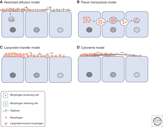
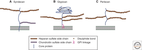
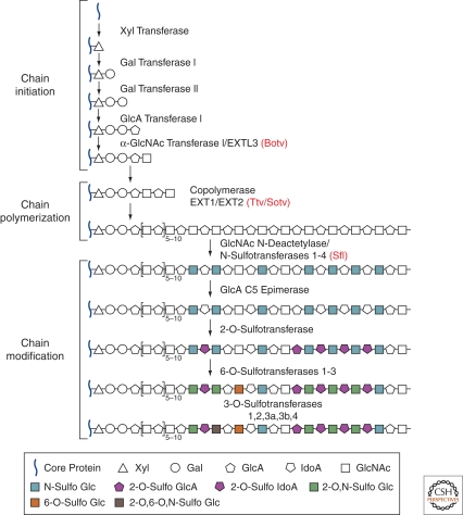
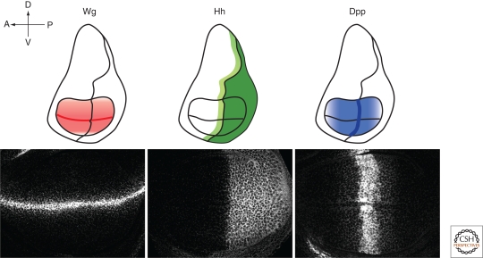
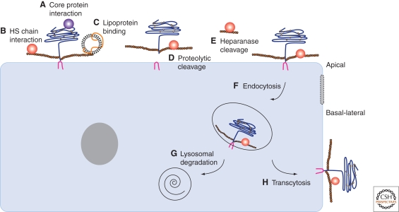
During development, secreted morphogens such as Wnt, Hedgehog (Hh), and BMP emit from their producing cells in a morphogenetic field, and specify different cell fates in a direct concentration-dependent manner. Understanding how morphogens form their concentration gradients to pattern tissues has been a central issue in developmental biology. Various experimental studies from Drosophila have led to several models to explain the formation of morphogen gradients. Over the past decade, one of the main findings in this field is the characterization of heparan sulfate proteoglycan (HSPG) as an essential regulator for morphogen gradient formation. Genetic and cell biological studies have showed that HSPGs can regulate morphogen activities at various steps including control of morphogen movement, signaling, and intracellular trafficking. Here, we review these data, highlighting recent findings that reveal mechanistic roles of HSPGs in controlling morphogen gradient formation.


Figures

Figure 1.
Figure 1.
Four models of morphogen gradient formation. (A) A model for restricted diffusion. Morphogens diffuse extracellularly by interaction with extracellular matrix proteins such as HSPGs, as well as other cell surface receptors and coreceptors. (B) A model for planar transcytosis. Morphogens are actively transported through repeated rounds of endocytosis and resecretion in receiving cells. (C) A model for lipoprotein transfer. Lipid-modified morphogens are packed into lipoprotein particles and transported in these vesicles. (D) A model for cytonemes. Morphogen receiving cells extend long, actin-based filopodia from the apical surface, called cytonemes, toward the morphogen source.
Figure 2.
Figure 2.
The three main classes of cell-surface heparan sulfate proteoglycans. (A) Syndecan core proteins are transmembrane proteins that contain a highly conserved carboxy-terminal cytoplasmic domain. Heparan sulfate (HS) chains attach to serine residues distal from the plasma membrane. Some syndecans also contain chondroitin sulfate (CS) chain(s) that attaches to serine residue(s) near the membrane. (B) The glypican core proteins are disulphide-stabilized globular core proteins that are linked to the plasma membrane by a glycosylphosphatidylinositol (GPI) anchor. HS chains link to serine residues adjacent to the plasma membrane. (C) Perlecans are secreted HSPGs that carry HS chains.
Figure 3.
Figure 3.
Heparan sulfate chain biosynthesis. Heparan sulfate (HS) glycosaminoglycan (GAG) chains are synthesized on a core protein by the sequential action of individual glycosyltransferases and modification enzymes, in a three-step process involving chain initiation, polymerization, and modification. HS chain synthesis begins with the assembly of a linkage tetrasaccharide on serine residues in the core protein. This process is catalyzed by four enzymes (Gal transferase I-III and α-GlcNAc transferase I), which add individual sugar residues sequentially to the nonreducing end of the growing chain. After the assembly of the linkage region, one or more α-GlcNAc transferases add a single α1,4-linked GlcNAc unit to the chain, which initiates the HS polymerization process. HS chain polymerization then takes place by the addition of alternating GlcA and GlcNAc residues, which is catalyzed by the EXT family proteins. As the chain polymerizes, it undergoes a series of modifications that include GlcNAc N-deacetylation and N-sulfation, C5 epimerization of GlcA to IdoA, and variable O-sulfation at C2 of IdoA and GlcA, at C6 of GlcNAc and GlcNS units, and, occasionally, at C3 of GlcN residues. The HS GAG chains are ∼100 or more sugar units long and have numerous structural heterogeneities. Four Drosophila enzymes, including Botv, Ttv, Sotv, and Sfl, which are homologs of vertebrate EXTL3, EXT1, EXT2, and N-deacetylase/N-sulfotransferase, respectively, are highlighted in red. (Gal) galactose, (GlcNAc) N-acetylglucosamine, (GlcA) glucuronic acid, (GlcNS) N-sulfoglucosamine, (IdoA) iduronic acid.
Figure 4.
Figure 4.
Distribution of Wg, Hh, and Dpp morphogens in the Drosophila wing imaginal disc. Drosophila wing imaginal discs are subdivided into anterior (A)/posterior (P) and dorsal (D)/ventral (V) compartments. In a third instar larvae wing disc, Wg is expressed at the D/V border and acts as a long-range morphogen to organize D/V patterning. Hh is expressed in the P compartment and moves in the A compartment to activate gene expression in a stripe of cells adjacent to the A/P compartment boundary. Dpp acts as a long-range morphogen that controls the growth and patterning of wing cells along the A/P axis beyond the central domain.
Figure 5.
Figure 5.
Regulations of cell-surface heparan sulfate proteoglycans. HSPGs can interact with morphogens directly either through their core proteins (A) or HS GAG chains (B). HSPGs also recruit lipoprotein particles by HS GAG chains (C). Proteolytic processing leads to the shedding of cell-surface HSPGs from the membrane (D), and heparanase cleaves the HS GAG chains, releasing bound morphogens (E). Cell-surface HSPGs are actively taken up by endocytosis (F), and then targeted to lysosome degradation (G) or transported from apical membrane to basal-lateral membrane by transcytosis (H).

Similar articles

Cited by

References

    1. Affolter M, Basler K 2007. The Decapentaplegic morphogen gradient: From pattern formation to growth regulation. Nat Rev Genet 8:663–674 - PubMed
    1. Ahn J, Ludecke HJ, Lindow S, Horton WA, Lee B, Wagner MJ, Horsthemke B, Wells DE 1995. Cloning of the putative tumour suppressor gene for hereditary multiple exostoses (EXT1). Nat Gen 11:137–143 - PubMed
    1. Akiyama T, Kamimura K, Firkus C, Takeo S, Shimmi O, Nakato H 2008. Dally regulates Dpp morphogen gradient formation by stabilizing Dpp on the cell surface. Dev Biol 313:408–419 - PMC - PubMed
    1. Baeg GH, Lin X, Khare N, Baumgartner S, Perrimon N 2001. Heparan sulfate proteoglycans are critical for the organization of the extracellular distribution of Wingless. Development 128:87–94 - PubMed
    1. Baeg GH, Selva EM, Goodman RM, Dasgupta R, Perrimon N 2004. The Wingless morphogen gradient is established by the cooperative action of Frizzled and Heparan Sulfate Proteoglycan receptors. Dev Biol 276:89–100 - PubMed

Publication types

LinkOut - more resources