Review
. 2009 Nov;1(5):a000497.
doi: 10.1101/cshperspect.a000497.

Symmetry breaking in plants: molecular mechanisms regulating asymmetric cell divisions in Arabidopsis

Affiliations
Review

Symmetry breaking in plants: molecular mechanisms regulating asymmetric cell divisions in Arabidopsis

Jalean J Petricka et al. Cold Spring Harb Perspect Biol. 2009 Nov.

Abstract

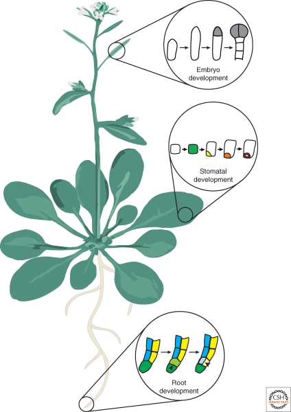
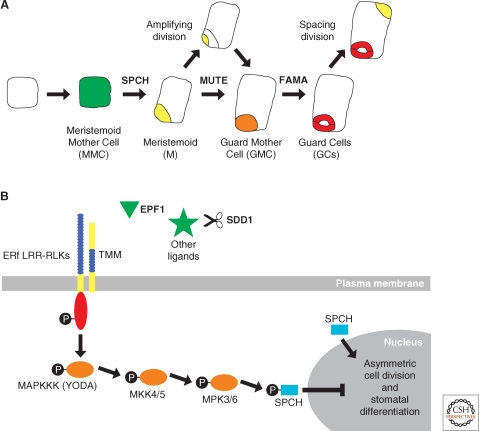
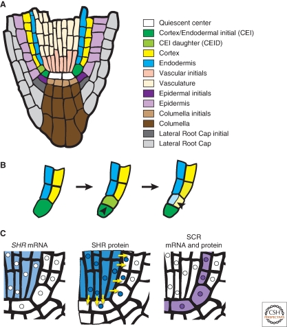
Asymmetric cell division generates cell types with different specialized functions or fates. This type of division is critical to the overall cellular organization and development of many multicellular organisms. In plants, regulated asymmetric cell divisions are of particular importance because cell migration does not occur. The influence of extrinsic cues on asymmetric cell division in plants is well documented. Recently, candidate intrinsic factors have been identified and links between intrinsic and extrinsic components are beginning to be elucidated. A novel mechanism in breaking symmetry was revealed that involves the movement of typically intrinsic factors between plant cells. As we learn more about the regulation of asymmetric cell divisions in plants, we can begin to reflect on the similarities and differences between the strategies used by plants and animals. Focusing on the underlying molecular mechanisms, this article describes three selected cases of symmetry-breaking events in the model plant Arabidopsis thaliana. These examples occur in early embryogenesis, stomatal development, and ground tissue formation in the root.


Figures

Figure 1.
Figure 1.
An Arabidopsis plant. Schematic of an adult plant depicting roots, leaves, stems, and flowers. In this article, three symmetry-breaking events are discussed. These examples are taken from different organs of the plant. The first example is taken from embryo development, which occurs after fertilization of the flower in the silique (seed pod) in Arabidopsis. Next, symmetry breaking in the leaf epidermis during stomatal development is discussed. Finally, the asymmetric cell division that gives rise to the ground tissue in the root is considered.
Figure 2.
Figure 2.
Embryo development and the asymmetric localization of factors in the embryo. (A) Schematic of embryo development focusing on the events from the egg to the eight-cell stage. After fertilization, the zygote expands and then divides asymmetrically to produce the apical and basal cells. Embryo stages are based on the number of cells in the apical domain only, thus the first zygotic division results in a one-cell stage embryo. The apical cell undergoes a series of divisions to generate the eight-celled proembryo. The basal cell undergoes a series of strictly transverse divisions to generate the suspensor. Later in development, the uppermost cell of the suspensor, the hypophysis, is incorporated into the embryo. Therefore, by the eight-cell stage, four anatomically distinct cell types are present: The upper (green) and lower (yellow-green) tiers of the proembryo, the hypophysis (cream), and the suspensor (white). The suspensor remains extraembryonic, whereas the remaining cell types give rise to the cotyledons, hypocotyl, and root of the mature embryo as depicted by the corresponding color scheme in the eight-cell stage and mature embryo. (B) Schematic of the WOX gene expression patterns. By the eight-cell stage, the expression domains of the WOX genes coincide with the four distinct cell types present. (C) Schematic of the auxin maxima and PIN7 localization. PIN7 localization on the upper membrane of basal cells directs the flow of auxin from basal to apical cells. This polarized movement of auxin generates an auxin maximum in the apical domain as determined by expression of the DR5 reporter.
Figure 3.
Figure 3.
Asymmetric cell division in stomatal development. (A) Schematic of a cell progressing through stomatal development. (A, from left to right) An undifferentiated leaf epidermal cell in Arabidopsis acquires Meristemoid Mother Cell fate (MMC, green) before undergoing an asymmetric cell division to produce a Meristemoid (M, yellow) and a larger sister cell (white). The M cell may then go through a series of asymmetric cell divisions, called amplifying divisions, before differentiating into a Guard Mother Cell (GMC, orange). The GMC divides symmetrically to produce a pair of Guard Cells (GCs, red) that together comprise a stomate. This process may reiterate when the larger sister cell divides asymmetrically in a spacing division to produce another M cell that is separated from existing stomata by one cell. The intrinsic factors SPCH, MUTE, and FAMA act sequentially to regulate asymmetric cell division and stomatal development. (B) Extrinsic factors and the signaling pathway proposed to negatively regulate stomatal development. First, a ligand binds to the leucine-rich repeats (LRR, blue) of a putative heterodimer complex between TMM and one of the ERf LRR-RLKs, which possess an intracellular kinase domain (red). This interaction is thought to initiate a cascade of phosphorylation events (black circles) involving downstream MAPK signaling proteins (orange), including YODA. Candidate ligands (green) acting to trigger this signaling cascade include small peptides such as EPF1 and putative unknown proteins that may be processed by SDD1. Biochemical evidence for this model is lacking, except for the final phosphorylation of SPCH by MPK3 and MPK6. SPCH phosphorylation ultimately results in repression of asymmetric cell division and stomatal differentiation. In contrast, when SPCH is unphosphorylated, it promotes asymmetric cell division and meristemoid fate, as shown in (A).
Figure 4.
Figure 4.
Symmetry breaking in ground tissue formation in the root. (A) Schematic of a longitudinal section of the Arabidopsis root tip with each cell type differentially colored. (B) Magnification of (A) focusing on the asymmetric cell divisions that generate the two cell layers of the ground tissue. (B, left) The CEI expands and then (B, center) divides transversely to regenerate itself and produce the CEID. (B, right) The CEID then divides longitudinally to generate the cells of the endodermis and cortex. (C) Schematic representation of the localization of SHR mRNA, SHR protein and SCR mRNA and protein. Yellow arrows depict the movement of SHR protein from the vasculature into the quiescent center, CEI, and endodermis. Note that SHR and SCR proteins are colocalized in the nuclei of these cell types. Nuclei are represented by small circles within the cells.

References

    1. Alessa L, Kropf DL 1999. F-actin marks the rhizoid pole in living Pelvetia compressa zygotes. Development 126:201–209 - PubMed
    1. Atchley WR, Fitch WM 1997. A natural classification of the basic helix-loop-helix class of transcription factors. Proc Natl Acad Sci 94:5172–5176 - PMC - PubMed
    1. Balasubramanian MK, Bi E, Glotzer M 2004. Comparative analysis of cytokinesis in budding yeast, fission yeast and animal cells. Curr Biol 14:R806–818 - PubMed
    1. Benfey PN, Linstead PJ, Roberts K, Schiefelbein JW, Hauser MT, Aeschbacher RA 1993. Root development in Arabidopsis: Four mutants with dramatically altered root morphogenesis. Development 119:57–70 - PubMed
    1. Berger D, Altmann T 2000. A subtilisin-like serine protease involved in the regulation of stomatal density and distribution in Arabidopsis thaliana. Genes Dev 14:1119–1131 - PMC - PubMed

Publication types

LinkOut - more resources