. 2010 Apr;459(5):689-703.
doi: 10.1007/s00424-009-0776-6. Epub 2010 Jan 12.

PPARbeta activation inhibits melanoma cell proliferation involving repression of the Wilms' tumour suppressor WT1

Affiliations

PPARbeta activation inhibits melanoma cell proliferation involving repression of the Wilms' tumour suppressor WT1

Jean-François Michiels et al. Pflugers Arch. 2010 Apr.

Abstract

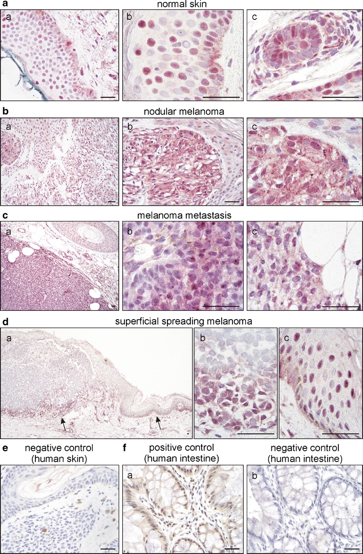
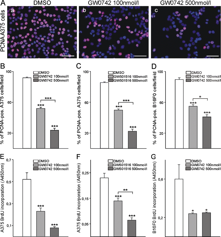
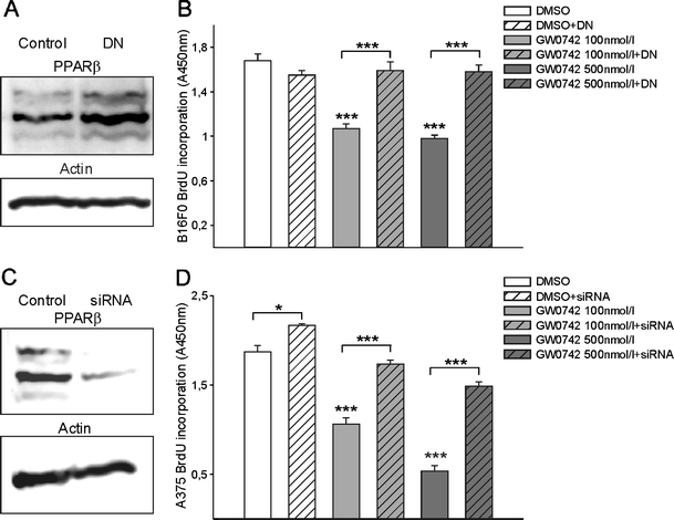
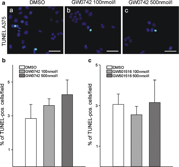
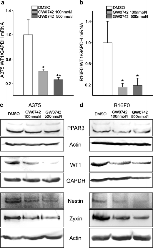
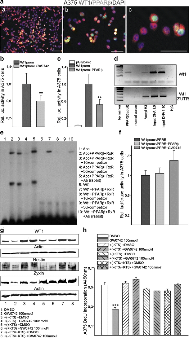
Peroxisome proliferator-activated receptors (PPARs) are ligand-activated transcription factors that strongly influence molecular signalling in normal and cancer cells. Although increasing evidence suggests a role of PPARs in skin carcinogenesis, only expression of PPARgamma has been investigated in human melanoma tissues. Activation of PPARalpha has been shown to inhibit the metastatic potential, whereas stimulation of PPARgamma decreased melanoma cell proliferation. We show here that the third member of the PPAR family, PPARbeta/delta is expressed in human melanoma samples. Specific pharmacological activation of PPARbeta using GW0742 or GW501516 in low concentrations inhibits proliferation of human and murine melanoma cells. Inhibition of proliferation is accompanied by decreased expression of the Wilms' tumour suppressor 1 (WT1), which is implicated in melanoma proliferation. We demonstrate that PPARbeta directly represses WT1 as (1) PPARbeta activation represses WT1 promoter activity; (2) in chromatin immunoprecipitation and electrophoretic mobility shift assays, we identified a binding element for PPARbeta in the WT1 promoter; (3) deletion of this binding element abolishes repression by PPARbeta and (4) the WT1 downstream molecules nestin and zyxin are down-regulated upon PPARbeta activation. Our findings elucidate a novel mechanism of signalling by ligands of PPARbeta, which leads to suppression of melanoma cell growth through direct repression of WT1.


Figures

Fig. 1
Fig. 1
PPARβ expression in normal skin, primary and metastatic melanoma. Representative examples of normal skin (a), nodular melanoma (b), a melanoma metastasis (c) and superficial spreading melanoma (d) stained for PPARβ (rabbit polyclonal antibody and VIP as substrate, purple). Sections were counterstained with haematoxylin to visualise nuclei. Note the mostly nuclear expression of PPARβ in keratinocytes, melanocytes and hair follicles and the heterogenous, both nuclear and cytoplasmatic expression in tumoural melanocytic lesions. Within the same melanoma metastasis, regions of moderate (c (b)) and low (c (c)) PPARβ expression coexist. In superficial spreading melanoma, PPARβ expression dominated in the invasive front of the tumour (d). Arrows in (d (a)) indicate the position of the high-power magnifications of the invasive front (d, (b)) and adjacent tissue (epidermis) with melanocyte atypia (d (c)). No staining could be observed by replacing the first antibody with normal serum (e). Human colon sections served as additional positive (f (a)) and negative (f (b)) controls (DAB substrate, brown). Scale bars indicate 50 µm
Fig. 2
Fig. 2
Pharmacological PPARβ activation in melanoma cell lines of human and murine origin. A375 (human) and B16F0 (mouse) melanoma cells were treated with different concentrations of two PPARβ agonists, GW0742, or GW501516, for 24 h. PPARβ activation inhibits melanoma cell proliferation. PPARβ-agonist-treated cells were immunostained with an anti-proliferating cell nuclear antigen (PCNA) antibody and counterstained with DAPI (a (ac)). Cells in seven random optical fields were counted and the percentage of PCNA-positive cells determined (bd; for each cell line and each agonist, n = 3, P < 0.001). Alternatively (eg), cells were incubated with 5-bromodeoxyuridine (BrdU) followed by immunological detection of the incorporated BrdU (for each cell line and each agonist, n = 4, P < 0.001 for A375 cells and, P < 0.05 for B16F0 cells)
Fig. 3
Fig. 3
Retroviral transduction of B16F0 melanoma cells with a dominant negative isoform of PPARβ (PPARβDN) and siRNA transfection of A375 melanoma cells against PPARβ and subsequent treatment with a PPARβ agonist GW0742. Western blot for PPARβ from lysates of B16F0 cells without or with retroviral transduction of the PPARβDN (a). Note the increase in PPARβ in the transduced cells reflecting the level of the transduced dominant negative PPARβ expression. β-actin served as standard. Cells with or without retroviral transduction with the PPARβDN, were treated with the PPARβ agonist GW0742 for 24 h. Note that the strong growth inhibitory effect of GW0742 was completely abolished in the cells expressing the dominant negative isoform of PPARβ (n = 4, P < 0.001; b). Western blot from lysates of A375 cells transfected with siRNA constructs against human PPARβ (c). Silenced and control cells were treated with the PPARβ agonist GW0742 for 24 h. Silencing of PPARβ restores proliferation in the presence of different concentrations of the agonist (n = 8; d)
Fig. 4
Fig. 4
Apoptosis assay of melanoma cells after pharmacological PPARβ activation. TdT-dUTP terminal nick-end labelling (TUNEL) labelling of A375 melanoma cells was performed after 24 h of treatment with either GW0742 or GW501516. Nuclei were counterstained with DAPI (a). Cells in ten random fields were counted and the percentage of TUNEL-positive cells determined (b, c; n = 4)
Fig. 5
Fig. 5
Pharmacological PPARβ activation decreases WT1 expression levels in melanoma cells. Quantitative reverse transcription (RT)-PCR for WT1 in A375 (a) and B16F0 (b) melanoma cells treated with GW0742 for 24 h. WT1 expression was normalised to GAPDH (n = 8, P < 0.01, P < 0.05). Western blot for PPARβ, WT1, nestin and zyxin from lysates of A375 (c) and B16F0 (d) melanoma cells treated with GW0742. Note that PPARβ expression remained stable upon pharmacological PPARβ activation, whereas WT1, nestin and zyxin expression was decreased. GAPDH and β-actin served as standards (n = 8 each)
Fig. 6
Fig. 6
PPARβ and WT1 co-localise partially in melanoma. In normal skin (a, b), no signal for WT1 could be observed in immunofluorescence double-labelling. Partial co-localisation (yellow) of PPARβ (Cy2, green) and WT1 (Cy3, red) could be detected in melanoma (cf). Nuclei were counterstained with DAPI (blue). Scale bars indicate 50 µm
Fig. 7
Fig. 7
PPARβ represses WT1. Co-localisation of PPARβ (Cy2, green) and WT1 (Cy3, red) in A375 melanoma cells (a). Nuclei were counterstained with DAPI (blue). Scale bars indicate 50 µm. b Transient transfection of the WT1 promoter in a luciferase vector in the presence of vehicle or 200 nM GW0742 in A375 cells. Luciferase activities were normalised for the activity of co-transfected β-galactosidase (n = 8, P < 0.01). c Transient co-transfections of a WT1 promoter construct together with a PPARβ expression construct in A375 melanoma cells (n = 12, P < 0.01). d Chromatin immunoprecipitation to analyse PPARβ protein interaction with WT1 regulatory elements. PPARβ protein binds to the WT1 promoter (upper panel), but not to the 3’-untranslated region (UTR) of WT1 (lower panel). Input DNA and immunoprecipitates obtained with acetylated histone 3 antibody served as positive controls. Negative controls were performed with normal serum instead of specific antibodies and with DNase-free water for the PCR. e Electrophoretic mobility shift assay demonstrating binding of the PPARβ/RXRα complex to the predicted consensus element of the WT1 promoter (lane 7). Unlabelled oligonucleotide from the acyl-CoA oxidase gene in the indicated molar excess was used as competitor (lanes 3, 4, 8 and 9). Supershift assays were performed by incubating the binding reactions with a polyclonal anti-PPARβ antibody (Ab rabbit, lanes 5 and 10). An oligonucleotide from the acyl-CoA oxidase gene served as positive control [1]. f Transient co-transfections of a WT1 construct with deletion of the identified 26 bp consensus motif together with PPARβ expression constructs or in the presence of 200 nM GW0742 in A375 melanoma cells (n = 12). Note that the 26 bp deletion abolished transactivation of the WT1 promoter by PPARβ or GW0742. g Western blot for WT1, nestin and zyxin from lysates of A375 cells transiently transfected with the WT1 (−KTS) or (+KTS) variant or a combination of both isoforms in a 50:50% ratio. Cells were subsequently treated with vehicle or 100 nM GW0742. Note that the decrease of WT1 expression by pharmacological PPARβ activation is abolished in the WT1 over-expressing cells. β-actin served as standard. h BrdU incorporation assay of the WT1 over-expressing cells, without or with pharmacological PPARβ activation. The growth inhibitory effect of GW0742 is abolished in the cells over-expressing any of the WT1 splice variants (n = 4, P < 0.001)

Similar articles

Cited by

References

    1. Amri EZ, Bonino F, Ailhaud G, Abumrad NA, Grimaldi PA. Cloning of a protein that mediates transcriptional effects of fatty acids in preadipocytes. Homology to peroxisome proliferator-activated receptors. J Biol Chem. 1995;270:2367–2371. doi: 10.1074/jbc.270.5.2367. - DOI - PubMed
    1. Balch CM, Buzaid AC, Soong SJ, et al. Final version of the American Joint Committee on Cancer staging system for cutaneous melanoma. J Clin Oncol. 2001;19:3635–3648. - PubMed
    1. Bastie C, Luquet S, Holst D, Jehl-Pietri C, Grimaldi PA. Alterations of peroxisome proliferator-activated receptor delta activity affect fatty acid-controlled adipose differentiation. J Biol Chem. 2000;275:38768–38773. doi: 10.1074/jbc.M006450200. - DOI - PubMed
    1. Berwick M, Wiggins C. The current epidemiology of cutaneous malignant melanoma. Front Biosci. 2006;11:1244–1254. doi: 10.2741/1877. - DOI - PubMed
    1. Burkart EM, Sambandam N, Han X, et al. Nuclear receptors PPARbeta/delta and PPARalpha direct distinct metabolic regulatory programs in the mouse heart. J Clin Invest. 2007;117:3930–3939. - PMC - PubMed

Publication types