Review
. 2010 Jun;67(11):1755-78.
doi: 10.1007/s00018-009-0238-5. Epub 2010 Jan 12.

Inositol trisphosphate 3-kinases: focus on immune and neuronal signaling

Affiliations
Review

Inositol trisphosphate 3-kinases: focus on immune and neuronal signaling

Michael J Schell. Cell Mol Life Sci. 2010 Jun.

Abstract

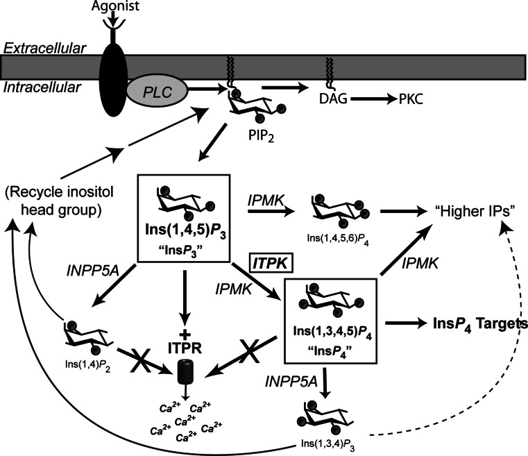
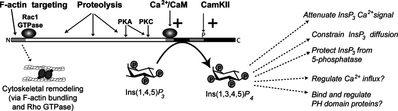
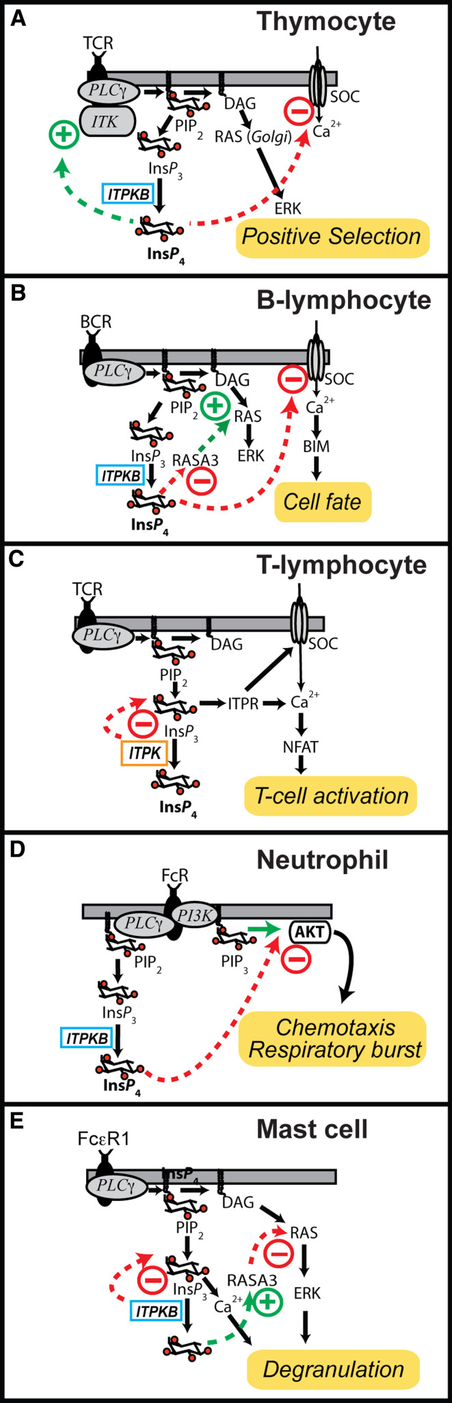
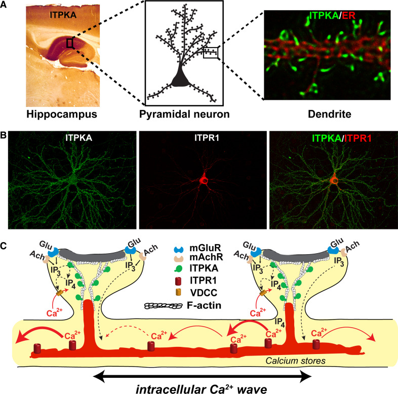
The localized control of second messenger levels sculpts dynamic and persistent changes in cell physiology and structure. Inositol trisphosphate [Ins(1,4,5)P(3)] 3-kinases (ITPKs) phosphorylate the intracellular second messenger Ins(1,4,5)P(3). These enzymes terminate the signal to release Ca(2+) from the endoplasmic reticulum and produce the messenger inositol tetrakisphosphate [Ins(1,3,4,5)P(4)]. Independent of their enzymatic activity, ITPKs regulate the microstructure of the actin cytoskeleton. The immune phenotypes of ITPK knockout mice raise new questions about how ITPKs control inositol phosphate lifetimes within spatial and temporal domains during lymphocyte maturation. The intense concentration of ITPK on actin inside the dendritic spines of pyramidal neurons suggests a role in signal integration and structural plasticity in the dendrite, and mice lacking neuronal ITPK exhibit memory deficits. Thus, the molecular and anatomical features of ITPKs allow them to regulate the spatiotemporal properties of intracellular signals, leading to the formation of persistent molecular memories.


Figures

Fig. 1
Fig. 1
Ins(1,4,5)P 3 signaling and metabolism. Activation of cell surface receptors triggers the canonical bifurcating pathway in which PLC hydrolyzes the lipid phosphatidylinositol 4,5 bisphosphate (PIP2). The soluble Ins(1,4,5)P 3 head group diffuses to the ER, where it gates the inositol trisphosphate receptor (ITPR), triggering Ca2+ release from intracellular stores. The diacylglycerol (DAG) remains in the lipid bilayer, where it activates protein kinase C (PKC). Ins(1,4,5)P 3 is subject to metabolism by cascades of phosphatases and kinases [10]. Inositol trisphosphate 3-kinases (ITPKs) phosphorylate Ins(1,4,5)P 3 at the 3-OH position to produce Ins(1,3,4,5)P 4, which does not gate ITPR channels but can bind various other protein targets in cells. Inositol polyphosphate multikinase (IPMK) phosphorylates Ins(1,4,5)P 3 twice, at the 6- and 3-positions, and these reactions govern the production of higher inositol phosphates (IPs) [34]. Both Ins(1,4,5)P 3 and Ins(1,3,4,5)P 4 are substrates for the type 1 inositol 5-phosphatase (INPP5A) [14]. If Ins(1,4,5)P 3 is the substrate, Ins(1,4,)P 2 is produced, which is dephosphorylated further and ultimately re-incorporated into new inositol lipids [17]. If Ins(1,3,4,5)P 4 is the substrate, Ins(1,3,4)P 3 is produced; the fate of Ins(1,3,4)P 3 varies among cells, and involves both kinase and phosphatase pathways [211]
Fig. 2
Fig. 2
Structure of ITPKs. a Comparison of crystal structures of kinase superfamily members. Crystal structures of human phosphatidylinositol 4-phosphate 5-kinase beta (PIPKIIβ, left) [212], yeast inositol polyphosphate multikinase (IPMK, middle) [27], and human ITPKA (right) [21]. Structures are oriented such that one is looking into the active sites, which bind ATP (red) through a conserved motif (SLL…IDF…, colored purple/orange). Inositol phosphates (IPs), such Ins(1,4,5)P 3 (InsP 3, blue) are oriented through their interaction with the IP lobe (blue), which holds the IP so that phosphotransfer from ATP is facilitated. IPMK (middle) has a smaller IP lobe than ITPK, allowing it to accommodate multiple IP substrates; by contrast, the more elaborated IP lobe found in ITPKs (right) holds Ins(1,4,5)P 3 in an orientation that allows phosphorylation at the 3-position only. b Domain structure of the three human ITPK isoforms compared to fly and worm varieties. The catalytic domains of ITPKs are highly conserved and located in the C-terminal parts of the protein (top domain diagram). By contrast, the amino terminal domains are divergent, and govern selective targeting and regulation in different cells and tissues. All ITPK crystal structures solved thus far have been obtained from catalytic fragments (gray), which lack the N-terminal regulatory regions; the catalytic region contains all of the substrate binding sites for ATP (red) and InsP 3 (blue). In the presence of Ca2+, all three human isoforms bind calmodulin (CaM) in a Ca2+-dependent manner, via a conserved domain [45]. The three human isoforms of ITPK show diversity in their N-termini [8]. Isoforms A [177] and B [77] bind filamentous actin (F-actin), while isoform C shows a mixed cytosolic/nuclear localization [213]. Arrowheads are labeled with point mutations that can selectively destroy domain functions in ITPKA or ITPKB. The L34P mutation destroys F-actin binding in ITPKA [133], while L139P and L159P reduce F-actin binding in ITPKB [77]. The W167R mutation destroys CaM binding in ITPKA; analogous mutations behave similarly for ITPKB and C [45]. Arrowheads in the blue and gray areas point to two of the many mutations in the active site that destroy catalytic activity [21]. The fruit fly Drosophila melanogaster possesses two isoforms [42], one of which is positively regulated by calmodulin [45]. The nematode Caenorhabditis elegans posesses one ITPK gene, which is not regulated by CaM and shows diversity generated by alternative splicing at the 5′ end [39]
Fig. 3
Fig. 3
Regulation of ITPKs and its consequences. ITPK action is regulated by at least four mechanisms. All mammalian ITPKs are positively regulated by Ca2+/calmodulin; this creates a negative feedback loop on the Ins(1,4,5)P 3 signal to release Ca2+ via ITPR [45]. ITPKs are also regulated by various protein kinases, such as protein kinase C (PKC), protein kinase A (PKA), and Ca2+/CaM-activated protein kinase II (CamKII). Phosphorylation at some sites activates the enzyme, and at others inhibits it (see text). The amino terminal regions of ITPKA and ITPKB bind F-actin, which targets the enzymes near sites of Ins(1,4,5)P 3 generation. The ITPKA amino terminal-binding region [177] affects F-actin structure through a mechanism independent of ITPKA catalytic activity [179]. This mechanism has been linked to the Rho family GTPase Rac1 [132]. The F-actin remodeling involves cross-linking or bundling of the actin filaments, and controls the selective targeting of ITPKA to dendritic spines [133]. ITPKs are subject to the actions of proteases, which usually cleave between the N-terminal targeting region and the catalytic region. Protease cleavage of ITPKs changes enzymatic localization [74, 75], and can also affect the sensitivity to or substrate sites for protein kinases [70]. ITPK catalysis has many suggested functions in cells [10]. These can be subdivided into functions involving the attenuation of Ins(1,4,5)P 3 signals via restriction of those signals to spatial and temporal domains through the control of Ins(1,4,5)P 3 lifetime, and those which depend on the specific actions of the enzymatic product Ins(1,3,4,5)P4. Cells possess a variety of Ins(1,3,4,5)P 4-binding proteins. Some of these bind Ins(1,3,4,5)P 4 selectively via their pleckstrin homology (PH) domains; others are channel proteins whose gating or conductance properties change upon Ins(1,3,4,5)P 4 binding (see text)
Fig. 4
Fig. 4
Diversity of ITPK signaling in immune cells. a Mice with targeted disruption in the gene for ITPKB exhibit major deficits in thymocyte maturation, with very few thymocytes undergoing positive selection [51, 52]. Activation of the T cell receptor in thymocytes derived from these mice results in less MAP kinase activation than in controls. At least two molecular mechanisms have been proposed to explain the phenotype. In one mechanism, Ins(1,3,4,5)P 4 triggers a feed-forward mechanism, whereby it binds and activates the interleukin-2 inducible T cell tyrosine kinase (ITK) [123] (green arrow). Downstream targets of ITK, such as phospholipase C gamma (PLCγ1), are thus hypoactive in the knockouts. A second mechanism proposes that store-operated Ca2+ channels (SOCs) are negatively regulated by Ins(1,3,4,5)P 4 (red arrow). In this model, lack of Ins(1,3,4,5)P 4 causes excessive Ca2+ influx, and this leads to deficits in Ca2+ homeostasis and its associated signaling [117]. b B lymphocytes from ITPKB knockout are anergic and undergo excessive apoptosis. Knockout B cells exhibit enhanced Ca2+ influx through SOCs, and this is attributed to a loss of normal inhibition on SOC by Ins(1,3,4,5)P 4 [117, 121] (red arrow). A second model suggests that Ins(1,3,4,5)P 4 is a negative regulator RASA3, a Ras GTPase activating protein that binds Ins(1,3,4,5)P 4 with high affinity [110] (red arrow). In this model, lack of Ins(1,3,4,5)P 4 leads to excessive GTPase activity, thus attenuating Ras and its downstream targets [110]. c Proposed role for ITPK signaling in mature, circulating T lymphocytes based on polymorphisms in the ITPKC gene linked to Kawasaki disease, an autoimmune disorder [59]. This model posits a hyperactive Ins(1,4,5)P 3-triggered Ca2+ response following activation of the T cell receptor (TCR), leading to excessive activation of the phosphatase calcineurin, causing dephosphorylation and over activation of the transcription factor NFAT. The hyperactive NFAT would result in an inappropriately large immune response in the vascular and mucosal systems, accounting for the Kawasaki disease phenotype. In contrast to the other immune models, no gain-of-function role for Ins(1,3,4,5)P 4 is invoked. d ITPKB also plays widespread roles in the innate immune system [120]. In neutrophils, Ins(1,3,4,5)P 4 generated downstream of Fc receptor (FcR) activation is proposed to compete with the head group of the lipid phosphatidylinositol (3,4,5)P3 (PIP3) for binding to the tyrosine kinase AKT. Thus, neutrophils derived from ITPKB knockouts exhibit enhanced PIP3-regulated responses, such as chemotaxis and the respiratory burst [53]. e Yet another mechanism is suggested to explain ITPKB signaling in mast cells [109]. Here, Fc receptor epsilon 1 (FcεR1) regulates degranulation via two ITPKB-dependent processes. The first is a straightforward reduction of Ca2+ release via ITPKB attenuation of Ins(1,4,5)P 3 signals (red arrow). The second, in contrast to the mechanism depicted in panel B, involves a positive regulation of RASA3 and concomitant negative effect on Ras activation status. In either mechanism shown in panel e, lack of ITPKB would produce hyperactive degranulation
Fig. 5
Fig. 5
ITPKA-regulated signaling in dendritic spines. a Regional and cellular localization of ITPKA in brain. Antibody staining against ITPKA (left) shows that the highest levels of ITPKA protein occur in the synapse-rich neuropil of the CA1 region of hippocampus (left, dark brown stain), while much lower expression occurs in the adjacent CA3 region. ITPKA is enriched in large pyramidal neurons, in dendritic spines (cartoon, middle). Deconvolved microscopic images (right) of a dendrite from a hipocampal neuron, co-labeled for ITPKA (green) and the endoplasmic reticulum (ER, red). Note how ITPKA is localized to Y-shaped structures, which lie inside dendritic spines and are situated between the synapse and the ER [147]. b Comparison of the localization of ITPKA and ITPR1 in a cultured hippocampal neuron. The image on the left (green) depicts a neuron transfected with ITPKA, which is localized toward the distal dendritic processes, in postsynaptic zones. The middle image (red) depicts transfected ITPR1, which is localized in the proximal ER and envelops the nucleus. The overlay (right) illustrates the inverse gradients of ITPK, located at distal (synaptic) sites, and ITPR1, located more proximally. c Cartoon depicting the spatial distribution of Ca2+ signaling systems regulated by ITPKA. Ins(1,4,5)P 3 is generated in spines chiefly through the activation of class 1 metabotropic glutamate (Glu) receptors (mGluR), which couple to phospholipase C beta (PLCβ). In the ITPKA-rich CA1 region of hippocampus, the mGluR subtype is mGluR5. Muscarinic acetylcholine (Ach) receptor (mAchR) subtypes M1 and M3 also occur in pyramidal neurons, and they constitute a second prominent Ins(1,4,5)P 3-generating system. Ins(1,4,5)P 3 generated inside dendritic spines diffuses to reach ITPR1, located in ER in or near the spine. ITPKA, which is intensely concentrated on bundles of filamentous actin (F-actin) inside spines [133], lies between sites of Ins(1,4,5)P 3 generation and action. Thus, the enzymatic activity of ITPKA is positioned as a molecular gatekeeper for synaptic Ins(1,4,5)P 3 signals. The Ca2+ released from intracellular stores can trigger an intracellular Ca2+ wave, which is driven by Ca2+-triggered Ca2+ release and also by Ins(1,4,5)P 3 produced in adjacent spines and propagates bi-directionally inside the main dendrite [137, 165]. Ins(1,3,4,5)P 4, the enzymatic product, has a direct effect on Ca2+ influx across the spine plasma membrane through its ability to enhance the activity of voltage-dependent Ca2+ channels (VDCCs) [118, 119]

Similar articles

Cited by

References

    1. Kummerow C, Junker C, Kruse K, Rieger H, Quintana A, Hoth M. The immunological synapse controls local and global calcium signals in T lymphocytes. Immunol Rev. 2009;231:132–147. - PubMed
    1. Kennedy MB, Beale HC, Carlisle HJ, Washburn LR. Integration of biochemical signalling in spines. Nat Rev Neurosci. 2005;6:423–434. - PubMed
    1. Berridge MJ, Bootman MD, Roderick HL. Calcium signalling: dynamics, homeostasis and remodelling. Nat Rev Mol Cell Biol. 2003;4:517–529. - PubMed
    1. Neves SR, Tsokas P, Sarkar A, Grace EA, Rangamani P, Taubenfeld SM, Alberini CM, Schaff JC, Blitzer RD, Moraru II, Iyengar R. Cell shape and negative links in regulatory motifs together control spatial information flow in signaling networks. Cell. 2008;133:666–680. - PMC - PubMed
    1. Willoughby D, Cooper DM. Organization and Ca2+ regulation of adenylyl cyclases in cAMP microdomains. Physiol Rev. 2007;87:965–1010. - PubMed

Publication types

MeSH terms

Substances

LinkOut - more resources