. 2010 Mar;19(3):544-57.
doi: 10.1002/pro.332.

Reversibly bound chloride in the atrial natriuretic peptide receptor hormone-binding domain: possible allosteric regulation and a conserved structural motif for the chloride-binding site

Affiliations

Reversibly bound chloride in the atrial natriuretic peptide receptor hormone-binding domain: possible allosteric regulation and a conserved structural motif for the chloride-binding site

Haruo Ogawa et al. Protein Sci. 2010 Mar.

Abstract

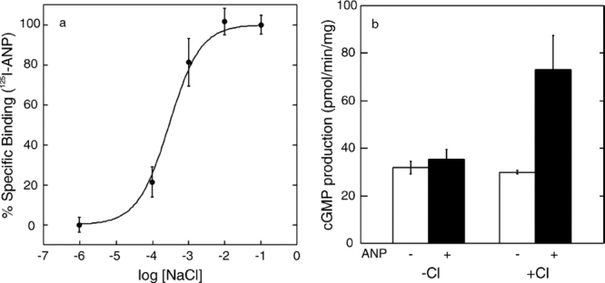
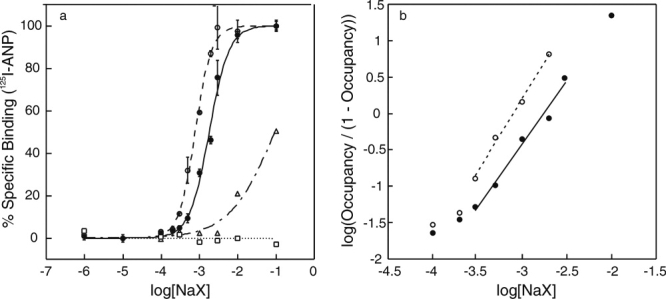
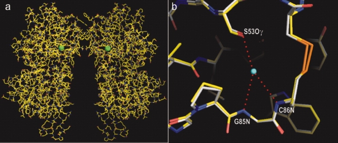
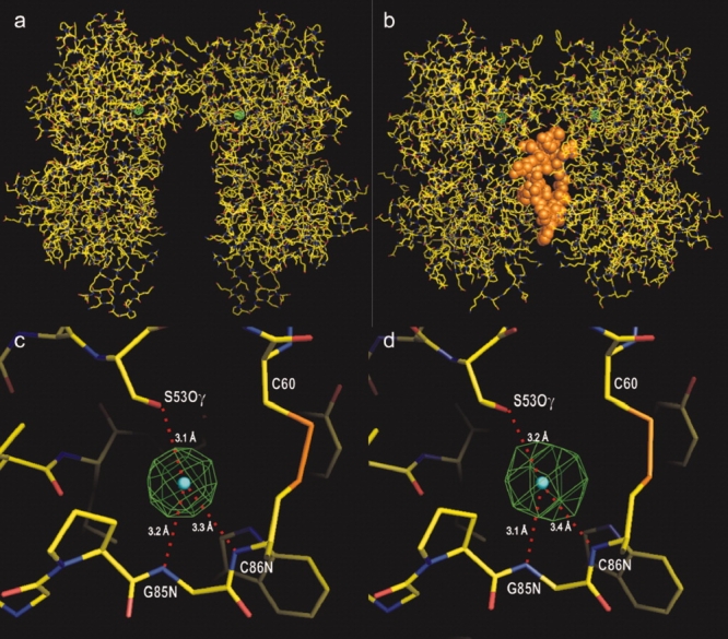
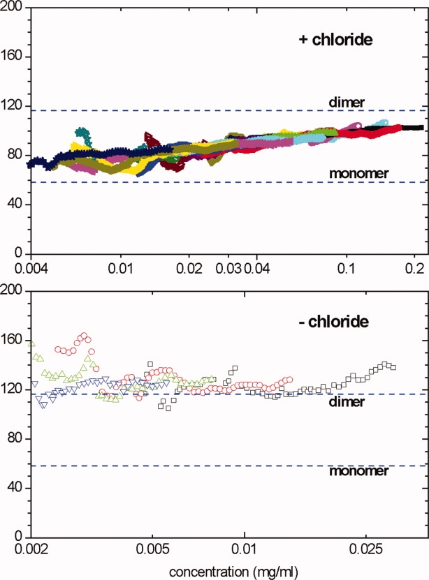
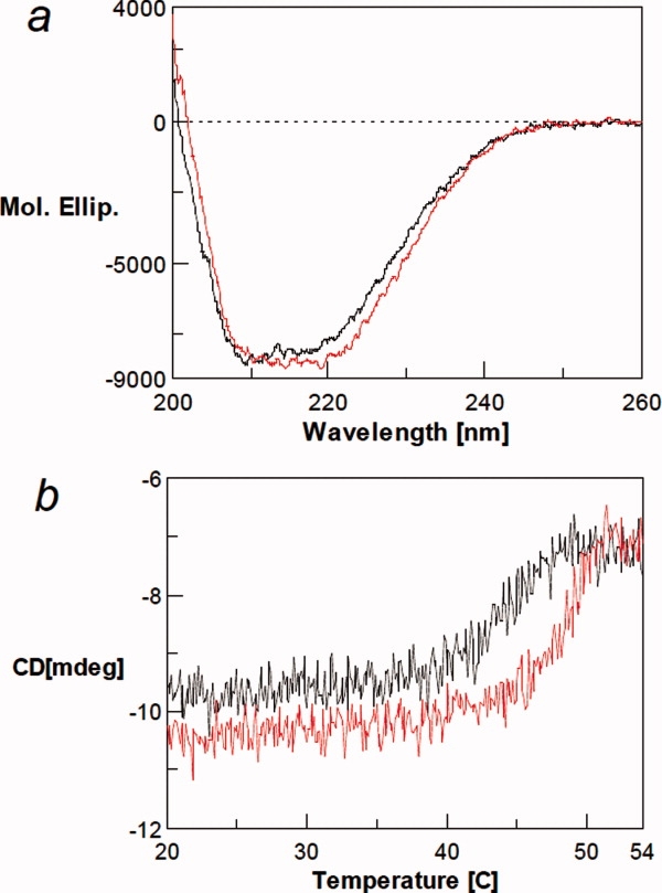
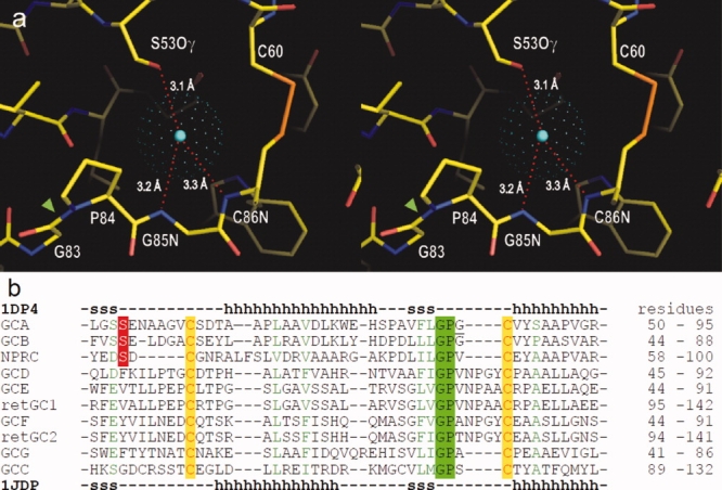
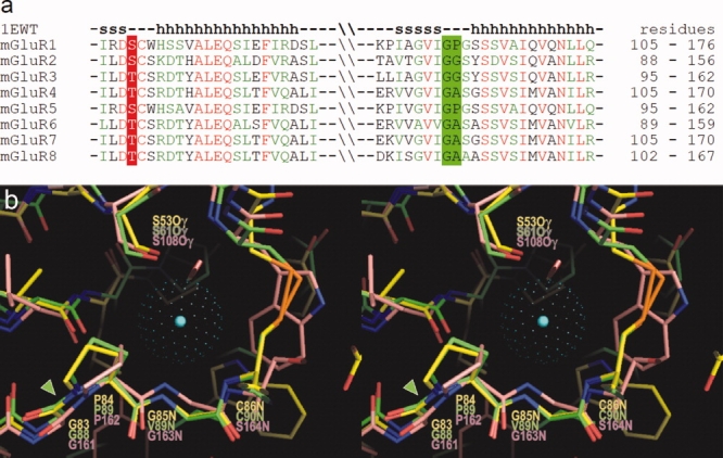
The binding of atrial natriuretic peptide (ANP) to its receptor requires chloride, and it is chloride concentration dependent. The extracellular domain (ECD) of the ANP receptor (ANPR) contains a chloride near the ANP-binding site, suggesting a possible regulatory role. The bound chloride, however, is completely buried in the polypeptide fold, and its functional role has remained unclear. Here, we have confirmed that chloride is necessary for ANP binding to the recombinant ECD or the full-length ANPR expressed in CHO cells. ECD without chloride (ECD(-)) did not bind ANP. Its binding activity was fully restored by bromide or chloride addition. A new X-ray structure of the bromide-bound ECD is essentially identical to that of the chloride-bound ECD. Furthermore, bromide atoms are localized at the same positions as chloride atoms both in the apo and in the ANP-bound structures, indicating exchangeable and reversible halide binding. Far-UV CD and thermal unfolding data show that ECD(-) largely retains the native structure. Sedimentation equilibrium in the absence of chloride shows that ECD(-) forms a strongly associated dimer, possibly preventing the structural rearrangement of the two monomers that is necessary for ANP binding. The primary and tertiary structures of the chloride-binding site in ANPR are highly conserved among receptor-guanylate cyclases and metabotropic glutamate receptors. The chloride-dependent ANP binding, reversible chloride binding, and the highly conserved chloride-binding site motif suggest a regulatory role for the receptor bound chloride. Chloride-dependent regulation of ANPR may operate in the kidney, modulating ANP-induced natriuresis.


Figures

Figure 1
Figure 1
(a) Chloride ion concentration dependence of ANP binding to the full-length ANPR in CHO cell membranes. (b) Stimulation of GCase activity by ANP (1 μM ANP) in the presence and absence of 100 mM NaCl. Bars represent the standard error of triplicate determinations.
Figure 2
Figure 2
ANP binding to ECD at varying halide concentrations. (a) Binding of 125I-ANP(4-28) to ECD was measured at varying concentrations of NaF (□), NaCl (•), NaBr (○), and NaI (Δ). Bars show the standard error in triplicate determinations. (b) Hill plots for chloride (•) and bromide (○) binding to ECD, in which the occupancy was calculated as the ANP-binding activity divided by the maximum activity at 100 mM halide concentration.
Figure 3
Figure 3
(a) Crystal structure of apo-ECD(Br) dimer determined by single-wavelength anomalous dispersion phasing. Bromide atoms are shown by green balls. (b) Close-up overlay view of the halide binding site in ECD(Br) (carbon atoms shown in white) and that in ECD(Cl) (carbon atoms in yellow). The center of the chloride atom is indicated by a blue dot. Hydrogen bonds are shown by red dotted lines. [Color figure can be viewed in the online issue, which is available at www.interscience.wiley.com.]
Figure 4
Figure 4
Anomalous difference Fourier map for bromide. (a and b) The electron density maps for bromine (in green) in apo-ECD(Br) and ANP-ECD(Br), respectively, are superimposed onto the apo-ECD(Cl) and ANP-ECD(Cl) structures. ANP is shown in orange. (c and d) Close-up views of the bromide density maps in a and b above, respectively. Bromide densities are contoured at 15 σ. The centers of the chloride atoms in apo-ECD(Cl) and ANP-ECD(Cl) are indicated by blue dots. [Color figure can be viewed in the online issue, which is available at www.interscience.wiley.com.]
Figure 5
Figure 5
Sedimentation equilibrium data for ECD in the presence (upper) or absence (lower) of 150 mM chloride. The local weight-average molecular mass within regions of the centrifuge cell was calculated from the slope of a ln(concentration) versus (radius)2/2 plot and plotted against the mean concentration for that region. Different data point styles and colors indicate different loading concentrations (see Materials and Methods section for details). The data in the upper include nine different loading concentrations at two rotor speeds; data collected at 9000 rpm are shown as open points, those at 13,000 rpm as filled points. The data in the lower are for four different loading concentrations at 13,000 rpm. [Color figure can be viewed in the online issue, which is available at www.interscience.wiley.com.]
Figure 6
Figure 6
(a) Far-UV CD spectra of ECD in the presence (red line) and absence of 100 mM NaCl (black line). (b) Thermal denaturation of ECD in the presence (red line) and absence of chloride ion (black line) as monitored by CD at 220 nm. Under the conditions used, there is little change in solvent signal with increasing temperature. [Color figure can be viewed in the online issue, which is available at www.interscience.wiley.com.]
Figure 7
Figure 7
Conserved structural motif for the chloride binding site. (a) Stereo view of the chloride binding site in apo-ECD (PDB 1DP4). The center of chloride atom is shown by a blue dot, and the van der Waals radius is presented by green dots. Chloride is hydrogen bonded to hydroxyl-group of Ser53, and backbone NH moieties of Gly85 and Cys86. The binding site also contains a single cis-peptide bond Gly83-Pro84 (green arrow head) in ECD. (b) Amino acid sequence alignment among natriuretic peptide receptors in the chloride-binding site region: ANPR or GCA; B-type natriuretic peptide receptor or GCB; NPCR; olfactory GCase GCD; eye retina GCases retGC, retGC2, GCE, and GCF; GCG; and guanylin/enterotoxin receptor or GCC. The sequences were aligned using the program ClustalW and are listed in the order of homology to GCA. The secondary structures in the X-ray structures of ANPR (1DP4) are shown above and below the aligned sequences (h, alpha-helix; s, beta-sheet). [Color figure can be viewed in the online issue, which is available at www.interscience.wiley.com.]
Figure 8
Figure 8
(a) Amino acid sequence alignment among mGluRs in the region homologous to the chloride binding site in ANPR. Residues in mGluRs, corresponding to Ser53 in ANPR, are conserved as either Ser or Thr. The Gly-Pro sequence in ANPR is conserved as Gly-Pro, Gly-Gly, or Gly-Ala. The secondary structures in mGluR1 are shown above the aligned sequences. (b) Overlay of the three-dimensional structures of the chloride binding sites in ANPR (PDB 1DP4) and NPCR (1JDP) and that of the water bound site in mGluR1 (1EWT) in stereo view. The carbon atoms in ANPR, NPCR, and mGluR1 are shown in yellow, green, and pink, respectively. The center of chloride atom in the ANPR is shown by a blue dot, and the van der Waals radius is presented by green dots. The conserved Gly-Pro cis-peptide bonds are highlighted by a green arrowhead. [Color figure can be viewed in the online issue, which is available at www.interscience.wiley.com.]

Similar articles

Cited by

References

    1. de Bold AJ, Borenstein HB, Veress AT, Sonnenberg H. A rapid and potent natriuretic response to intravenous injection of atrial myocardial extract in rats. Life Science. 1981;28:89–94. - PubMed
    1. Grammer RT, Fukumi H, Inagami T, Misono KS. Rat atrial natriuretic factor. Purification and vasorelaxant activity. Biochem Biophys Res Commun. 1983;116:696–703. - PubMed
    1. Currie MG, Geller DM, Cole BR, Boylan JG, YuSheng W, Holmberg SW, Needleman P. Bioactive cardiac substances: potent vasorelaxant activity in mammalian atria. Science. 1983;221:71–73. - PubMed
    1. Ogawa H, Qiu Y, Ogata CM, Misono KS. Crystal structure of hormone-bound atrial natriuretic peptide receptor extracellular domarotation mechanism for transmembrane signal transduction. J Biol Chem. 2004;279:28625–28631. - PubMed
    1. Misono KS, Ogawa H, Qiu Y, Ogata CM. Structural studies of the natriuretic peptide receptor: a novel hormone-induced rotation mechanism for transmembrane signal transduction. Peptides. 2005;26:957–968. - PubMed

Publication types

Substances

Associated data

LinkOut - more resources