

Review
Dental enamel: genes define biomechanics
Affiliations
- PMID: 20066874
- PMCID: PMC2825347
Item in Clipboard
Review
Dental enamel: genes define biomechanics
J Calif Dent Assoc.
2009 Dec.
Abstract
Regulated gene expression assembles an extracellular proteinaceous matrix to control biomineralization and the resultant biomechanical function of tooth enamel. The importance of the dominant enamel matrix protein, amelogenin (Amel); a minor transiently expressed protein, dentin sialoprotein (Dsp); an electrogenic sodium bicarbonate cotransporter (NBCe1); the timely removal of the proteinaceous matrix by a serine protease, Kallikrein-4 (Klk4); and the late-stage expression of Amelotin (Amtn) on enamel biomechanical function were demonstrated and measured using mouse models.
Figures
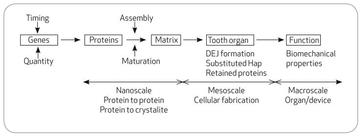
The hierarchy of tooth formation from genes to functioning teeth
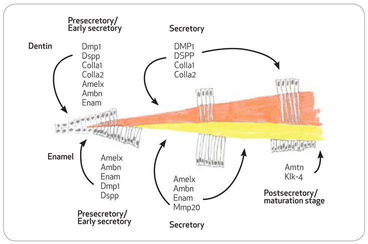
Gene expression through enamel and dentin formation. Schematic of proteins secreted into the dentin matrix by odontoblasts and the enamel matrix by ameloblasts at the various stages of formation. With the exception of collagen, odontoblasts, and ameloblasts, expression profiles for the secreted proteins is very similar in the very early secretory stages. During the secretory stages both odontoblasts and ameloblasts gene expression profiles are entirely distinct as the enamel matures, amelotin, and kallikrein-4 are upregulated.
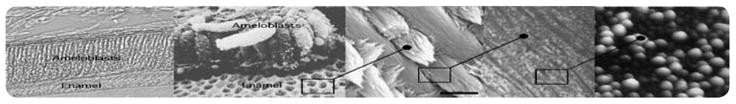
Enamel formation. From left to right: A layer of columnar ameloblasts lay down their protein matrix, which becomes mineralized to form enamel. Each individual ameloblast produces a cylindrical matrix field that becomes mineralized as a rod within a “honeycomb”-like continuum of interrod; the complex migratory vectors of ameloblasts weave rods into a complex fibrous network. Each rod is composed of multitudes of individual crystallites organized by amelogenin nanospheres; amelogenin proteins spontaneously form nanospheres in physologic conditions.
References
-
- Paine ML, White SN, et al. Regulated gene expression dictates enamel structure and tooth function. Matrix Biology. 2001;20(56):273–92. - PubMed
-
- Barlett JD, Ganss B, et al. Protein-protein interactions of the developing enamel matrix. Curr Top Devel Biol. 2006;74:57–115. - PubMed
-
- White SN, Luo W, et al. Biological organization of hydroxyapatite crystallites into a fibrous continuum toughens and controls anisotropy in human enamel. J Dent Res. 2001;80(1):321–6. - PubMed
-
- Snead ML, Paine ML, et al. Transgene animal model for protein expression and accumulation into forming enamel. Conn Tiss Res. 1998;38(14):279–86. - PubMed
-
- Kubota K, Lee DH, et al. Fluoride induces ER stress in ameloblasts responsible for dental enamel formation. J Biol Chem. 2005;280(24):23194–202. - PubMed
Publication types
MeSH terms
Substances
Grants and funding
LinkOut - more resources
Full Text Sources
Miscellaneous