Review
. 2010 Feb;45(1):50-69.
doi: 10.3109/10409230903505596.

Integrating prokaryotes and eukaryotes: DNA transposases in light of structure

Affiliations
Review

Integrating prokaryotes and eukaryotes: DNA transposases in light of structure

Alison Burgess Hickman et al. Crit Rev Biochem Mol Biol. 2010 Feb.

Abstract

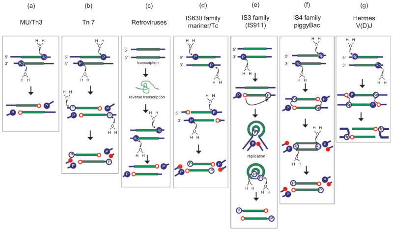
DNA rearrangements are important in genome function and evolution. Genetic material can be rearranged inadvertently during processes such as DNA repair, or can be moved in a controlled manner by enzymes specifically dedicated to the task. DNA transposases comprise one class of such enzymes. These move DNA segments known as transposons to new locations, without the need for sequence homology between transposon and target site. Several biochemically distinct pathways have evolved for DNA transposition, and genetic and biochemical studies have provided valuable insights into many of these. However, structural information on transposases - particularly with DNA substrates - has proven elusive in most cases. On the other hand, large-scale genome sequencing projects have led to an explosion in the number of annotated prokaryotic and eukaryotic mobile elements. Here, we briefly review biochemical and mechanistic aspects of DNA transposition, and propose that integrating sequence information with structural information using bioinformatics tools such as secondary structure prediction and protein threading can lead not only to an additional level of understanding but possibly also to testable hypotheses regarding transposition mechanisms. Detailed understanding of transposition pathways is a prerequisite for the long-term goal of exploiting DNA transposons as genetic tools and as a basis for genetic medical applications.


Conflict of interest statement

Declaration of interest

The authors report no conflicts of interest. The authors alone are responsible for the content and writing of the paper.

Figures

Figure 1
Figure 1
Dealing with the second strand. The color code is as follows: transposon DNA (green); flanking donor DNA (blue); target phosphates destined to be removed from the final liberated transposon (filled blue circles with a white “P”); phosphates destined to remain as 5′ transposon ends (open blue circles); the preferred stereoisomer, Sp or Rp, where known, is indicated within the circles; liberated 3′OH groups involved in strand joining reactions (open red circles); 3′OH destined to be removed from the liberated transposon (filled red circles); H2O is the attacking nucleophile in the hydrolysis reactions. (a) The Mu and Tn3 cleavage reactions. Note that the preferred stereoisomer has been demonstrated only for Mu and not for Tn3. (b) Tn7 cleavage reactions. Cleavage of the transferred strand (top of panel) is shown occurring prior to cleavage of the non-transferred strand (middle) leading to liberation of the transposon from flanking donor DNA (bottom of panel), although this order of cleavage reactions has not been demonstrated experimentally. The two types of cleavage are catalyzed by different enzymes. (c) Retroviral “processing” reaction, equivalent to cleavage of the transferred strand. An initial transcription step from the integrated provirus is indicated. The RNA genome is then encapsidated with a second copy and undergoes reverse transcription following infection to generate the double strand DNA integration intermediate. The intermediate is flanked by only short fragments of donor material and does not require second strand processing for insertion. (d) Transposition by the members of the IS630 family and the Tc1/Mariner superfamily is initiated by cleavage of the non-transferred strand (top of panel) at several bases within the transposon end (middle) leaving these bases attached to the liberated flanks following cleavage of the transferred strand (bottom). (e) For IS911, IS2, IS3 and other members of the IS3 family, single-end hydrolysis occurs (top). The liberated 3′OH then directs a strand transfer reaction to the same strand several bases 5′ to the other end of the element. This results in the formation of a single-strand circle which is then resolved into a transposon circle by replication from the free 3′OH (filled red circle). Single-strand hydrolysis at each 3′ end within the circle generates a linear transposon which can then undergo integration. (f ) The IS4 family and piggyBac have similar mechanisms. Following initial nucleophilic attack on the Rp target phosphate, the liberated 3′OH attacks an Sp phosphate in a trans-strand transfer reaction to generate a hairpin intermediate, liberating the transposon from its flanking donor DNA and inverting the target phosphate to its Rp configuration. These then become the substrates for a second hydrolysis. Note that the stereochemistry has been analyzed only in the case of Tn10. (g) Hermes and V(D)J transposition occur by initial cleavage of the non-transferred strand (top). The liberated 3′OH on the donor flank then attacks the opposite strand (middle) to generate hairpin structure on the donor flank (bottom). The stereochemistry has been analyzed for V(D)J only. Modified and reprinted from Turlan and Chandler (2000), with permission from Elsevier.
Figure 2
Figure 2
Ribbon diagrams of the aligned catalytic cores of four DNA transposases and of HIV-1 integrase. Residues shown in orange are the carboxylate active side residues, in green are the W residues of the Tn5 transposase and Hermes that are important in the reactions, in blue are the YRK residues of the YREK motif and in yellow is W298 of the Tn5 transposase. The insertion domains of the Tn5 transposase and Hermes are shown in red (note that there is a 15 amino acid loop from residues S481 to K496 that is disordered – and therefore not visible – in Hermes). The proteins are drawn to scale.
Figure 3
Figure 3
Ribbon diagram of the catalytic core domain of HIV-1 integrase, with the standard secondary structure elements highlighted.

Similar articles

Cited by

References

    1. Allingham JS, Wardle SJ, Haniford DB. Determinants for hairpin formation in Tn10 transposition. EMBO J. 2001;20:2931–2942. - PMC - PubMed
    1. Andrews JD, Gloor GB. A role for the KP leucine zipper in regulating P element transposition in Drosophila melanogaster. Genet. 1995;141:587–594. - PMC - PubMed
    1. Ariyoshi M, Vassylyev DG, Iwasaki H, Nakamura H, Shinagawa H, Morikawa K. Atomic structure of the RuvC resolvase: A Holliday junction-specific endonuclease from E. coli. Cell. 1994;78:1063–1072. - PubMed
    1. Babu MM, Iyer LM, Balaji S, Aravind L. The natural history of the WRKY-GCM1 zinc fingers and the relationship between transcription factors and transposons. Nucl Acids Res. 2006;34:6505–6520. - PMC - PubMed
    1. Bailin T, Mo XM, Sadofsky MJ. A RAG1 and RAG2 tetramer complex is active in cleavage in V(D)J recombination. Mol Cell Biol. 1999;19:4664–4671. - PMC - PubMed

Publication types

LinkOut - more resources