. 2010 Jan;20(1):33-41.
doi: 10.1089/thy.2009.0296.

Metastatic phenotype is regulated by estrogen in thyroid cells

Affiliations

Metastatic phenotype is regulated by estrogen in thyroid cells

Shilpi Rajoria et al. Thyroid. 2010 Jan.

Abstract

Background: Over 200 million people worldwide are affected by thyroid proliferative diseases, including cancer, adenoma, and goiter, annually. The incidences of thyroid malignancies are three to four times higher in women, suggesting the possible involvement of estrogen. Based on this observed sex bias, we hypothesize that estrogen modulates the growth and metastatic propensity of thyroid cancer cells.

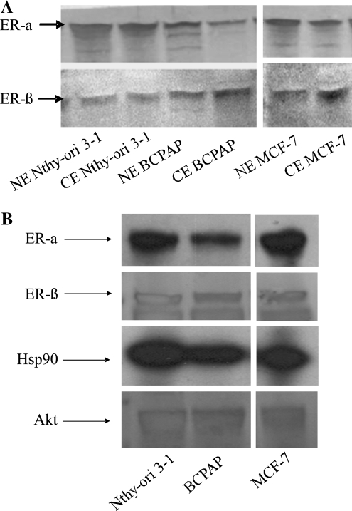
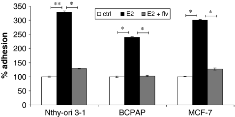
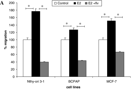
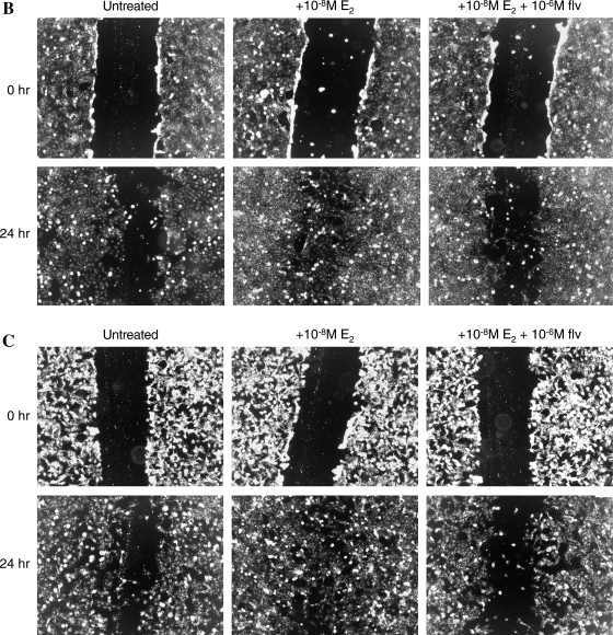
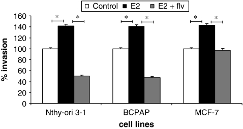
Methods: In this study, two thyroid cell lines (Nthy-ori 3-1 and BCPAP) were evaluated for the presence of estrogen receptor (ER) by Western blot analysis and estrogen responsiveness by using a cell proliferation assay. In addition, the effect of estradiol (E(2)) on modulation of metastatic phenotype was determined by using in vitro adhesion, migration, and invasion assays.

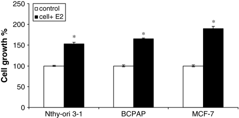
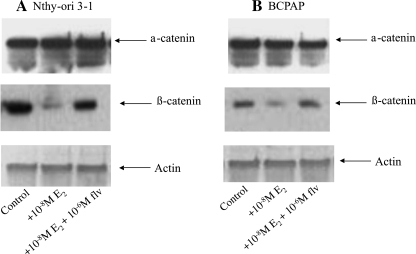
Results: Thyroid cells expressed a functionally active ER-alpha and ER-beta as evidenced by 50-150% enhancement of proliferation in the presence of E(2). E(2) also enhanced adhesion, migration, and invasion of thyroid cells in an in vitro experimental model system that, based on our results, is modulated by beta-catenin.

Conclusion: Our data provide evidence that the higher incidence of thyroid cancer in women is potentially attributed to the presence of a functional ER that participates in cellular processes contributing to enhanced mitogenic, migratory, and invasive properties of thyroid cells. These findings will enable and foster the possible development of antiestrogenic therapy targeting invasion and migration, thus affecting metastatic propensity.


Figures

FIG. 1.
FIG. 1.
Estrogen stimulates proliferation of thyroid cells. Effect of E2 on proliferation of Nthy-ori 3-1 and BCPAP was determined by the trypan blue dye exclusion assay. Cells were seeded at a density of 1 × 105 cells per well and allowed to adhere overnight. The starvation medium (10% charcoal-dextran-treated FBS) was then added for 24 hours, followed by stimulation with 10−8 M E2 for 24 hours. MCF-7, estrogen-responsive human breast cancer cells were used as a positive control. Cell growth is expressed as percent increase in viable cells + E2 (black bars) relative to untreated control (white bars). The asterisk denotes statistically significant increase (p < 0.05) in the experimental groups compared with controls. E2, estradiol; FBS, fetal bovine serum.
FIG. 2.
FIG. 2.
(A) Thyroid cells express estrogen receptor (ER). Nuclear (NE) and cytoplasmic (CE) protein fractions (15 μg) were resolved by sodium dodecyl sulfate–polyacrylamide gel electrophoresis, and Western blot analysis, for ER-α (45 KDa) and ER-β (60 KDa), was performed. MCF-7, ER-positive cells were used as positive control. (B) ER is complexed with Akt and HSP90 in thyroid cells. Cell lysates were subjected to immunoprecipitation with antibodies to human ER-α and ER-β followed by Western blot analysis for ER-α, ER-β, HSP90, and Akt, respectively (top to bottom panel of immunoprecipitation and Western blot). MCF-7 (ER positive) was used as a positive control for detection of Hsp90 and Akt complex with ER.
FIG. 3.
FIG. 3.
Estrogen stimulates adhesion of thyroid cells. About 5 × 105 cells were plated in six-well culture dishes in the presence of ±10−8 M E2 ± fulvestrant. After 2.5 hours, the medium was removed, and wells were gently washed with phosphate-buffered saline to remove nonadherent cells. Viable adhered cells were removed by scraping and counted using trypan blue dye exclusion test. The groups are as follows: untreated (white bars), E2 treated (black bars), and E2 + fulvestrant treated (flv) (gray bars). MCF-7 was used as a positive control. E2-mediated increase in the number of adhesive cells expressed as % of control (100%). * denotes statistically significant differences (p < 0.05) between experimental and control groups. ** denotes statistically significant differences (p < 0.001) between experimental and control groups.
FIG. 4.
FIG. 4.
Estrogen enhances the migration of thyroid cells. (A) About 2.5 × 104 cells were resuspended in 500 μL of Rosswell Park Memorial Institute (RPMI) with 1% FBS ± E2 and fulvestrant and seeded on upper chamber of BD Biocoat Control Inserts (8-μm pore membrane filters). Seven hundred and fifty microliters of Rosswell Park Memorial Institute (RPMI) containing 5% FBS was added to the bottom chamber as chemoattractant. After 18 hours, cells that migrated and adhered on the lower surface of the membrane were fixed, stained, and counted in 10 × field. The groups are as follows: untreated (white bars), E2 treated (black bars), and E2 + fulvestrant treated (gray bars). MCF-7 cells were used as a positive control for migration studies. Data are expressed as numbers of cells counted (migrated cells) per 10 × field micrograph for each sample well and normalized to the untreated control. The asterisk denotes statistically significant differences (p < 0.05) between experimental and control groups. (B) Scratch wound assay for Nthy-ori 3-1. (C) Scratch wound assay for BCPAP. About 5 × 105 cells were plated and were allowed to grow to semiconfluent cell monolayers when three vertical wounds were caused per well using a pipette tip, and the cells were allowed to migrate in the presence of E2 and/or fulvestrant. The cells were observed under 5 × field every 3 hours until the cells completely migrated from one end of scratch to other end.
FIG. 4.
FIG. 4.
Estrogen enhances the migration of thyroid cells. (A) About 2.5 × 104 cells were resuspended in 500 μL of Rosswell Park Memorial Institute (RPMI) with 1% FBS ± E2 and fulvestrant and seeded on upper chamber of BD Biocoat Control Inserts (8-μm pore membrane filters). Seven hundred and fifty microliters of Rosswell Park Memorial Institute (RPMI) containing 5% FBS was added to the bottom chamber as chemoattractant. After 18 hours, cells that migrated and adhered on the lower surface of the membrane were fixed, stained, and counted in 10 × field. The groups are as follows: untreated (white bars), E2 treated (black bars), and E2 + fulvestrant treated (gray bars). MCF-7 cells were used as a positive control for migration studies. Data are expressed as numbers of cells counted (migrated cells) per 10 × field micrograph for each sample well and normalized to the untreated control. The asterisk denotes statistically significant differences (p < 0.05) between experimental and control groups. (B) Scratch wound assay for Nthy-ori 3-1. (C) Scratch wound assay for BCPAP. About 5 × 105 cells were plated and were allowed to grow to semiconfluent cell monolayers when three vertical wounds were caused per well using a pipette tip, and the cells were allowed to migrate in the presence of E2 and/or fulvestrant. The cells were observed under 5 × field every 3 hours until the cells completely migrated from one end of scratch to other end.
FIG. 5.
FIG. 5.
Estrogen enhances invasion of thyroid cells. Matrigel-coated invasion chambers (8-μm pore membrane filters) were used for invasion assay. About 2.5 × 104 cells per insert were resuspended in 500 μL medium containing 1% FBS ± 10−8 M E2 and/or 10−6 M fulvestrant were transferred onto the upper surface of filters in the chamber. Seven hundred and fifty microliters of growth medium containing 5% FBS was used as chemoattractant. Invasion was calculated based on the percent of cells invading through the growth-factor-reduced Matrigel invasion chambers relative to the cells migrating through control membrane after 18 hours when counted under 10 × field. The groups are as follows: untreated (white bars), E2 treated (black bars), and E2 + fulvestrant treated (gray bars). MCF-7 was used as a positive control for invasion. Data are represented as percent invasion (according to manufacturer's protocol), which is mean number of invaded cells per 10 × field micrograph for each sample well relative to the migration through the control membrane and normalized to the untreated control. * denotes statistically significant differences (p < 0.05) between experimental and control groups.
FIG. 6.
FIG. 6.
E2 affects metastasis via downregulation of β-catenin. Nthy-ori 3-1 (A) and BCPAP (B) cells were treated with E2 ± fulvestrant. Whole cell protein (20 μg) was resolved by sodium dodecyl sulfate–polyacrylamide gel electrophoresis followed by Western blot analysis for α-catenin, β-catenin, and actin as a protein loading control.

References

    1. Hodgson NC. Button J. Solorzano CC. Thyroid cancer: is the incidence still increasing? Ann Surg Oncol. 2004;11:1093–1097. - PubMed
    1. Jemal A. Siegel R. Ward E. Hao Y. Xu J. Thun MJ. Cancer statistics, 2009. CA Cancer J Clin. 2009;59:225–249. - PubMed
    1. Vasko VV. Saji M. Molecular mechanisms involved in differentiated thyroid cancer invasion and metastasis. Curr Opin Oncol. 2007;19:11–17. - PubMed
    1. Mackenzie EJ. Mortimer RH. Thyroid nodules and thyroid cancer. Med J Aust. 2004;180:242–247. - PubMed
    1. Kondo T. Ezzat S. Asa SL. Pathogenetic mechanisms in thyroid follicular-cell neoplasia. Nat Rev Cancer. 2006;6:292–306. - PubMed

Publication types

MeSH terms