

Impact of degradable macromer content in a poly(ethylene glycol) hydrogel on neural cell metabolic activity, redox state, proliferation, and differentiation
- PMID: 20067398
- PMCID: PMC2949233
- DOI: 10.1089/ten.TEA.2009.0509
Impact of degradable macromer content in a poly(ethylene glycol) hydrogel on neural cell metabolic activity, redox state, proliferation, and differentiation
Abstract
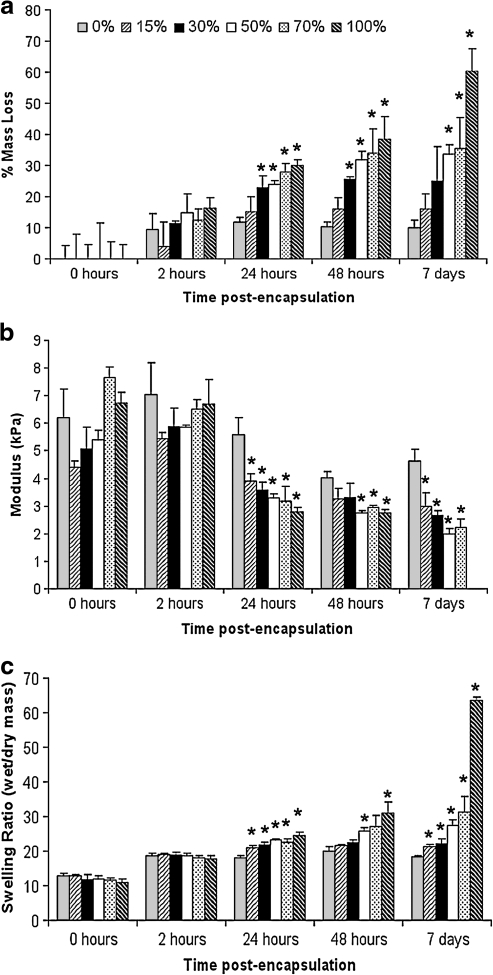
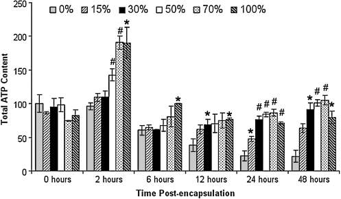
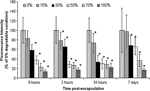
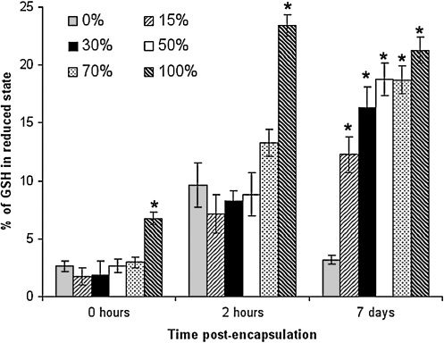
Hydrogels that degrade at different rates were prepared by copolymerizing slowly degrading macromer poly(ethylene glycol) (PEG) dimethacrylate with a faster degrading macromer poly(lactic acid)-b-PEG-b-poly(lactic acid) dimethacrylate. A clinically relevant population of neural cells composed of differentiated neurons and multipotent precursor cells was cultured within hydrogels. Within 2 h after encapsulation, metabolic activity was higher in hydrogels prepared with increasing levels of degradable content. This improvement was accompanied by a reduction in intracellular redox state and an increase in the fraction of glutathione in the reduced state, both of which persisted throughout 7 days of culture and which may be the result of radical scavenging by lactic acid. Importantly, an increase in cellular proliferation was observed in gels prepared with increasing degradable macromer content after 7 days of growth without a shift in the cellular composition of the culture toward the glial cell phenotype. The findings of this study provide additional insight into the growth of neural cells in PEG-based hydrogels. Results suggest that lactic acid released during gel degradation may impact the function of encapsulated cells, a finding of general interest to biomaterials scientists who focus on the development of degradable polymers for cell culture and drug delivery devices.
Figures

Similar articles
-
Control of neural cell composition in poly(ethylene glycol) hydrogel culture with soluble factors.Tissue Eng Part A. 2011 Nov;17(21-22):2805-15. doi: 10.1089/ten.tea.2010.0654. Epub 2011 Aug 8. Tissue Eng Part A. 2011. PMID: 21823990 Free PMC article.
-
Cell viability of chitosan-containing semi-interpenetrated hydrogels based on PCL-PEG-PCL diacrylate macromer.J Biomater Sci Polym Ed. 2005;16(3):301-16. doi: 10.1163/1568562053654149. J Biomater Sci Polym Ed. 2005. PMID: 15850286
-
Effect of macromer weight percent on neural cell growth in 2D and 3D nondegradable PEG hydrogel culture.J Biomed Mater Res A. 2010 Sep 15;94(4):1162-71. doi: 10.1002/jbm.a.32787. J Biomed Mater Res A. 2010. PMID: 20694983
-
Encapsulation and 3D culture of human adipose-derived stem cells in an in-situ crosslinked hybrid hydrogel composed of PEG-based hyperbranched copolymer and hyaluronic acid.Stem Cell Res Ther. 2013 Mar 21;4(2):32. doi: 10.1186/scrt182. Stem Cell Res Ther. 2013. PMID: 23517589 Free PMC article.
-
Poly(lactic acid) based hydrogels.Adv Drug Deliv Rev. 2016 Dec 15;107:192-205. doi: 10.1016/j.addr.2016.07.004. Epub 2016 Jul 16. Adv Drug Deliv Rev. 2016. PMID: 27432797 Review.
Cited by
-
Preparation of a Fucoidan-Grafted Hyaluronan Composite Hydrogel for the Induction of Osteoblast Differentiation in Osteoblast-Like Cells.Materials (Basel). 2021 Mar 2;14(5):1168. doi: 10.3390/ma14051168. Materials (Basel). 2021. PMID: 33801348 Free PMC article.
-
Microphysiological Systems for Neurodegenerative Diseases in Central Nervous System.Micromachines (Basel). 2020 Sep 16;11(9):855. doi: 10.3390/mi11090855. Micromachines (Basel). 2020. PMID: 32947879 Free PMC article. Review.
-
Building biocompatible hydrogels for tissue engineering of the brain and spinal cord.J Funct Biomater. 2012 Nov 15;3(4):839-63. doi: 10.3390/jfb3040839. J Funct Biomater. 2012. PMID: 24955749 Free PMC article.
-
Functional and Biomimetic Materials for Engineering of the Three-Dimensional Cell Microenvironment.Chem Rev. 2017 Oct 25;117(20):12764-12850. doi: 10.1021/acs.chemrev.7b00094. Epub 2017 Oct 9. Chem Rev. 2017. PMID: 28991456 Free PMC article. Review.
-
Control of neural cell composition in poly(ethylene glycol) hydrogel culture with soluble factors.Tissue Eng Part A. 2011 Nov;17(21-22):2805-15. doi: 10.1089/ten.tea.2010.0654. Epub 2011 Aug 8. Tissue Eng Part A. 2011. PMID: 21823990 Free PMC article.
References
-
- Kordower J.H. Freeman T.B. Snow B.J. Vingerhoets F.J.G. Mufson E.J. Sanberg P.R. Hauser R.A. Smith D.A. Nauert G.M. Perl D.P. Olanow C.W. Neuropathological evidence of graft-survival and striatal reinnervation after the transplantation of fetal mesencephalic tissue in a patient with parkinsons-disease. N Engl J Med. 1995;332:1118. - PubMed
-
- Clarkson E.D. Zawada W.M. Adams F.S. Bell K.P. Freed C.R. Strands of embryonic mesencephalic tissue show greater dopamine neuron survival and better behavioral improvement than cell suspensions after transplantation in parkinsonian rats. Brain Res. 1998;806:60. - PubMed
Publication types
MeSH terms
Substances
Grants and funding
LinkOut - more resources
Full Text Sources
Other Literature Sources
