

Insulin-like growth factor I messenger RNA and protein are expressed in the human lymph node and distinctly confined to subtypes of macrophages, antigen-presenting cells, lymphocytes and endothelial cells
- PMID: 20067534
- PMCID: PMC2770682
- DOI: 10.1111/j.1365-2567.2009.03136.x
Insulin-like growth factor I messenger RNA and protein are expressed in the human lymph node and distinctly confined to subtypes of macrophages, antigen-presenting cells, lymphocytes and endothelial cells
Abstract
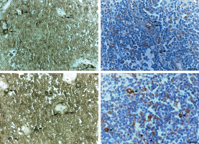
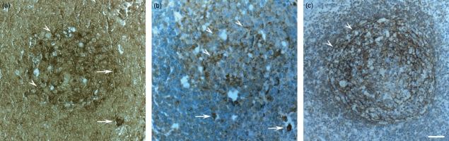
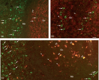
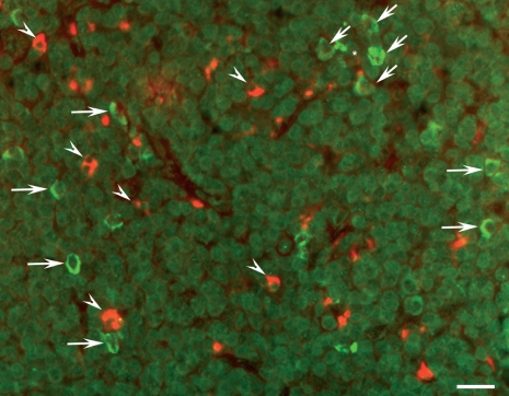
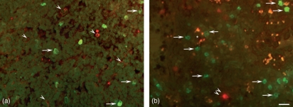
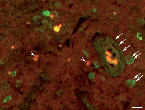
Insulin-like growth factor I (IGF-I) is a potent hormone that stimulates growth and differentiation and inhibits apoptosis in numerous tissues. Preliminary evidence suggests that IGF-I exerts differentiating, mitogenic and restoring activities in the immune system but the sites of synthesis of local IGF-I are unknown. Identification of these sites would allow the functional role of local IGF-I to be clarified. The presence of IGF-I in non-immune cells suggests that it acts as a trophic factor, while its occurrence in subtypes of lymphocytes or antigen-presenting cells indicates paracrine/autocrine direct regulatory involvement of IGF-I in the human immune response. The present study investigated the location of IGF-I messenger RNA and protein on archival human lymph node samples by in situ hybridization, immunohistochemistry and double immunofluorescence staining using an IGF-I probe and antisera specific for human IGF-I and CD3 (T lymphocytes), CD20 (B lymphocytes), CD68 (macrophages), CD21 (follicular dendritic cells), S100 (interdigitating dendritic cells) and podoplanin (fibroblastic reticular cells). Numerous cells within the B- and T-cell compartments expressed the IGF-I gene, and the majority of these cells were identified as macrophages. Solitary follicular dendritic cells exhibited IGF-I. A few T lymphocytes, and no B lymphocytes, contained IGF-I immunoreactive material. Furthermore, IGF-I immunoreactive cells outside the follicles that did not react with CD3, CD20, S100 or podoplanin markers were identified as high-endothelial venule (HEV) cells. From this we conclude that the main task of IGF-I in human non-tumoral lymph node may be autocrine and paracrine regulation of the differentiation, stimulation and survival of lymphocytes, antigen-presenting cells and macrophages and the differentiation and maintenance of HEV cells.
Figures
Similar articles
-
Immunophenotyping of Sheep Paraffin-Embedded Peripheral Lymph Nodes.Front Immunol. 2018 Dec 11;9:2892. doi: 10.3389/fimmu.2018.02892. eCollection 2018. Front Immunol. 2018. PMID: 30619264 Free PMC article.
-
IGF-I is distinctly located in the bony fish pituitary as revealed for Oreochromis niloticus, the Nile tilapia, using real-time RT-PCR, in situ hybridisation and immunohistochemistry.Gen Comp Endocrinol. 2007 Jan 1;150(1):87-95. doi: 10.1016/j.ygcen.2006.07.013. Epub 2006 Sep 11. Gen Comp Endocrinol. 2007. PMID: 16963049
-
Expression of the intercellular adhesion molecule-1 on high endothelial venules and on non-lymphoid antigen handling cells: interdigitating cells, antigen transporting cells and follicular dendritic cells.Cell Tissue Res. 1995 Jan;279(1):47-54. doi: 10.1007/BF00300690. Cell Tissue Res. 1995. PMID: 7895264
-
Cooperation of liver cells in health and disease.Adv Anat Embryol Cell Biol. 2001;161:III-XIII, 1-151. doi: 10.1007/978-3-642-56553-3. Adv Anat Embryol Cell Biol. 2001. PMID: 11729749 Review.
-
Insulin-like growth factors and insulin-like growth factor binding proteins in the endometrium. Effect of intrauterine levonorgestrel delivery.Hum Reprod. 2000 Aug;15 Suppl 3:173-81. doi: 10.1093/humrep/15.suppl_3.173. Hum Reprod. 2000. PMID: 11041233 Review.
Cited by
-
Individuals with isolated congenital GH deficiency due to a GHRH receptor gene mutation appear to cope better with SARS-CoV-2 infection than controls.Endocrine. 2021 May;72(2):349-355. doi: 10.1007/s12020-021-02728-8. Epub 2021 Apr 16. Endocrine. 2021. PMID: 33860882 Free PMC article.
-
Overload training inhibits phagocytosis and ROS generation of peritoneal macrophages: role of IGF-1 and MGF.Eur J Appl Physiol. 2013 Jan;113(1):117-25. doi: 10.1007/s00421-012-2418-5. Epub 2012 May 17. Eur J Appl Physiol. 2013. PMID: 22592456 Clinical Trial.
-
Macrophages From Subjects With Isolated GH/IGF-I Deficiency Due to a GHRH Receptor Gene Mutation Are Less Prone to Infection by Leishmania amazonensis.Front Cell Infect Microbiol. 2019 Aug 30;9:311. doi: 10.3389/fcimb.2019.00311. eCollection 2019. Front Cell Infect Microbiol. 2019. PMID: 31544067 Free PMC article.
-
Spleen: A new role for an old player?World J Gastroenterol. 2011 Sep 7;17(33):3776-84. doi: 10.3748/wjg.v17.i33.3776. World J Gastroenterol. 2011. PMID: 21987619 Free PMC article. Review.
-
The role of obesity in gastrointestinal cancer: evidence and opinion.Therap Adv Gastroenterol. 2014 Jan;7(1):38-50. doi: 10.1177/1756283X13501786. Therap Adv Gastroenterol. 2014. PMID: 24381646 Free PMC article. Review.
References
-
- Jones JI, Clemmons DR. Insulin-like growth factors and their binding proteins: biological actions. Endocr Rev. 1995;16:3–34. - PubMed
-
- Wallenius K, Sjögren K, Peng XD, et al. Liver-derived IGF-I regulates GH secretion at the pituitary level in mice. Endocrinology. 2001;142:4762–70. - PubMed
-
- Cohen P. Overview of the IGF-I system. Horm Res. 2006;65(Suppl. 1):3–8. - PubMed
-
- Reinecke M. Insulin and the insulin-like growth factors. In: Reinecke M, Zaccone G, Kapoor BG, editors. Fish Endocrinology. Vol. 1. Enfield (NH), Jersey, Plymouth, USA: Science Publishers; 2006. pp. 3–13.
-
- Reinecke M, Schmid AC, Heyberger-Meyer B, Hunziker EB, Zapf J. Effect of growth hormone and insulin-like growth factor I (IGF-I) on the expression of IGF-I messenger ribonucleic acid and peptide in rat tibial growth plate and articular chondrocytes in vivo. Endocrinology. 2000;141:2847–53. - PubMed
MeSH terms
Substances
LinkOut - more resources
Full Text Sources
