. 2010 Jan 12:9:5.
doi: 10.1186/1476-4598-9-5.

Smoothened as a new therapeutic target for human osteosarcoma

Affiliations

Smoothened as a new therapeutic target for human osteosarcoma

Masataka Hirotsu et al. Mol Cancer. .

Abstract

Background: The Hedgehog signaling pathway functions as an organizer in embryonic development. Recent studies have demonstrated constitutive activation of Hedgehog pathway in various types of malignancies. However, it remains unclear how Hedgehog pathway is involved in the pathogenesis of osteosarcoma. To explore the involvement of aberrant Hedgehog pathway in the pathogenesis of osteosarcoma, we investigated the expression and activation of Hedgehog pathway in osteosarcoma and examined the effect of SMOOTHENED (SMO) inhibition.

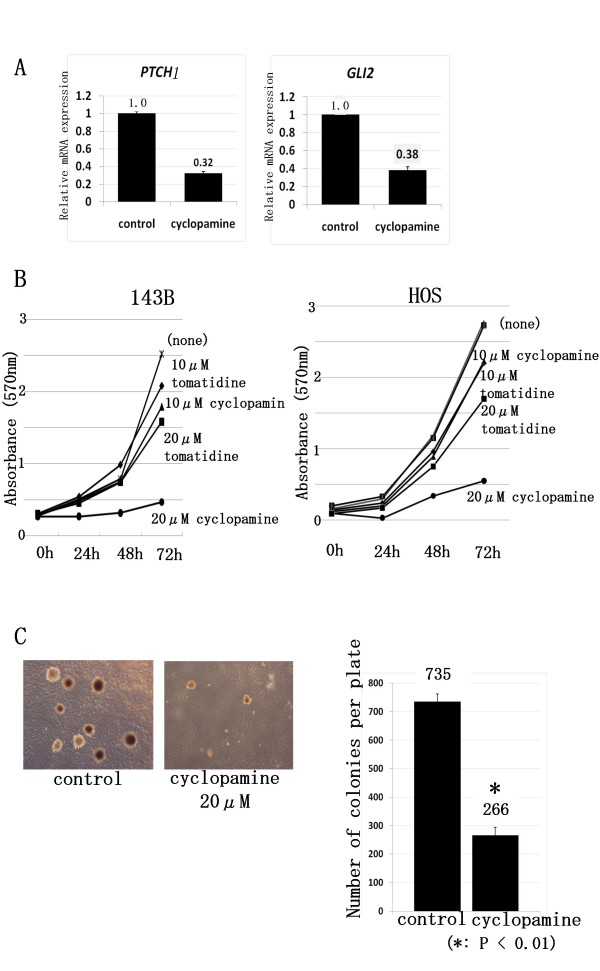
Results: To evaluate the expression of genes of Hedgehog pathway, we performed real-time PCR and immunohistochemistry using osteosarcoma cell lines and osteosarcoma biopsy specimens. To evaluate the effect of SMO inhibition, we did cell viability, colony formation, cell cycle in vitro and xenograft model in vivo. Real-time PCR revealed that osteosarcoma cell lines over-expressed Sonic hedgehog, Indian hedgehog, PTCH1, SMO, and GLI. Real-time PCR revealed over-expression of SMO, PTCH1, and GLI2 in osteosarcoma biopsy specimens. These findings showed that Hedgehog pathway is activated in osteosarcomas. Inhibition of SMO by cyclopamine, a specific inhibitor of SMO, slowed the growth of osteosarcoma in vitro. Cell cycle analysis revealed that cyclopamine promoted G1 arrest. Cyclopamine reduced the expression of accelerators of the cell cycle including cyclin D1, cyclin E1, SKP2, and pRb. On the other hand, p21(cip1) wprotein was up-regulated by cyclopamine treatment. In addition, knockdown of SMO by SMO shRNA prevents osteosarcoma growth in vitro and in vivo.

Conclusions: These findings suggest that inactivation of SMO may be a useful approach to the treatment of patients with osteosarcoma.


Figures

Figure 1
Figure 1
Expression of activated Hh-GLI pathway molecules. Total RNA extracted from osteosarcoma cell lines were used for real-time PCR. Real-time PCR revealed that 4 of 5 human osteosarcoma cell lines increased Sonic Hedgehog (SHH) 2.1- to 18.8-fold (Fig. 1). In addition, 5 of 5 osteosarcoma cell lines increased Desert Hedgehog 1.3- to 24.4-fold (Fig. 1). To further examine Hh pathway molecules expression, we performed real-time PCR for Hh receptors and Hh target genes. PTCH1 was up-regulated 2.7-to 65.8-fold in 5 of 5 human osteosarcoma cell lines. SMO was up-regulated 2.1-to 5.8-fold in 4 of 5 human osteosarcoma cell lines. SMO was up-regulated 2.1-to 5.8-fold in 4 of 5 human osteosarcoma cell lines. GLI1 was up-regulated 2.5-to 8.9-fold in 5 of 5 human osteosarcoma cell lines. GLI2 was up-regulated 1.2-to 9.9-fold in 5 of 5 human osteosarcoma cell lines. The comparative Ct (ΔΔCt) method was used to determine fold change in expression using βII-microglobulin, GAPDH or ACTB. Each sample was run minimally at three concentrations in triplicate (error bar means S.D.). The experiment was triplicate with similar results.
Figure 2
Figure 2
Activation of Hh pathway in patient' biopsy samples. Real-time PCR revealed that 9 of 9 human biopsy specimens of osteosarcoma increased SMO 1.44- to 55.5-fold. Real-time PCR revealed that expression of PTCH1 was increased in 8 of 9 patients' biopsy samples 2.44- to 29.4-fold. GLI2 was up-regulated 2.5-to 58.4-fold in 9 of 9 human biopsy specimens of osteosarcoma. The comparative Ct (ΔΔCt) method was used to determine fold change in expression using βII-microglobulin, ACTB, and GAPDH. Each sample was run minimally at three concentrations in triplicate (error bar means S.D.). The experiment was triplicate with similar results.
Figure 3
Figure 3
Inhibition of Hh pathway prevents osteosarcoma growth in vitro. A, We performed real-time PCR to determine which concentration of cyclopamine effectively inhibited Hh-GLI activity in osteosarcoma cells, and then measured the expression of the Hh-GLI pathway target PTCH1 and GLI2. Cyclopamine at 20 μM reduced mRNA levels of PTCH1 in 143B cell (error bar means S.D.). The comparative Ct (ΔΔCt) method was used to determine fold change in expression using ACTB. Each sample was run minimally at three concentrations in triplicate (error bar means S.D.). The experiment was triplicate with similar results. B, Growth of viable 143B and HOS cells over 3 days was slowed in dose-dependent fashion by cyclopamine treatment. The experiment was triplicate with similar results. C, Colony formation assay revealed cyclopamine reduced colony formation in soft agar. The experiment was triplicate with similar results. (*: P < 0.01) (error bar means S.D.)
Figure 4
Figure 4
Cyclopamine treatment promotes G1 arrest. A, HOS and143B cells were treated with 10 μM cyclopamine. After 48-hour treatment cells were collected and subjected to cell cycle analysis. When 143B cells were cultured without cyclopamine, 39.8% of cells were in G1 phase. On the other hand, when cultured with cyclopamine, 56.6% of cells were in G1 phase. In the case of HOS cells cultured without GSI, 55.4% of cells were in G1 phase, while 72.3% of cells were in G1 phase when treated with cyclopamine (error bar means S.D.). B, Real-time PCR was performed to quantify mRNAs of cell cycle related genes. Twenty-four-hour treatment with cyclopamine reduced levels of cyclin D1, Cyclin E1, SKP2, and NMYC transcription (error bar means S.D.). The comparative Ct (ΔΔCt) method was used to determine fold change in expression using βII-microglobulin and GAPDH. Each sample was run minimally at three concentrations in triplicate (error bar means S.D.). The experiment was triplicate with similar results. C, Western blot analysis of levels of cell cycle-related genes. Forty-eight-hour treatment with cyclopamine reduced levels of expression of cyclin D1, cyclin E1, SKP2, and phosphprylated RB (pRb) proteins. Expression of P21cip1 protein was upregulated by cyclopamine treatment. The experiment was triplicate with similar results (cyclopamine: 10 μM).
Figure 5
Figure 5
Knock down of SMO by SMO shRNA prevents osteosarcoma growth in vitro. A, Real-time PCR revealed that SMO shRNA effectively knock down SMO mRNA. (error bar means S.D.). The comparative Ct (ΔΔCt) method was used to determine fold change in expression using ACTB. Each sample was run minimally at three concentrations in triplicate (error bar means S.D.). The experiment was triplicate with similar results. B, Growth of viable 143B cells over 4 days was slowed by SMO shRNA. The experiment was triplicate with similar results. C, Colony formation assay revealed that SMO shRNA reduced colony formation in soft agar. The experiment was triplicate with similar results. (*: P < 0.01) (Error bar means S.D.)
Figure 6
Figure 6
SMO shRNA prevents osteosarcoma xenograft growth in vivo and prolongs survival. A, SMO shRNA or control shRNA transfected 143B cells (1 × 10⁶) were inoculated subcutaneously. Established 143B tumors were measured. The tumor volume at day 7 was set at 1, and tumor volumes at subsequent time points were calculated. SMO shRNA inhibited tumor growth at 8 weeks compared to control shRNA. B, Kaplan-Meier survival curves from SMO shRNA groups and control shRNA. Kaplan-Meier analysis showed that SMO shRNA conferred a significant survival benefit (n = 6, p < 0.05). C, Immunohistochemical examination of ki67 was performed in xenograft tumors. SMO shRNA decreased SMO RNA in vivo. Ki67 staining revealed that proliferation of osteosarcoma cells was decreased by GSI treatment. The number of Ki67-positive cells was decreased to 30% of control revel by SMO shRNA (error bar means S.D.) (*: P < 0.01).

Similar articles

Cited by

References

    1. Gibbs CP Jr, Weber K, Scarborough MT. Malignant bone tumors. Instr Course Lect. 2002;51:413–428. - PubMed
    1. Igney FH, Krammer PH. Death and anti-death: tumour resistance to apoptosis. Nat Rev Cancer. 2002;2:277–288. doi: 10.1038/nrc776. - DOI - PubMed
    1. Lafleur EA, Koshkina NV, Stewart J, Jia SF, Worth LL, Duan X, Kleinerman ES. Increased Fas expression reduces the metastatic potential of human osteosarcoma cells. Clin Cancer Res. 2004;10:8114–8119. doi: 10.1158/1078-0432.CCR-04-0353. - DOI - PubMed
    1. Horowitz JM, Park SH, Bogenmann E, Cheng JC, Yandell DW, Kaye FJ, Minna JD, Dryja TP, Weinberg RA. Frequent inactivation of the retinoblastoma anti-oncogene is restricted to a subset of human tumor cells. Proc Natl Acad Sci USA. 1990;87:2775–2779. doi: 10.1073/pnas.87.7.2775. - DOI - PMC - PubMed
    1. Ingham PW, McMahon AP. Hedgehog signaling in animal development: paradigms and principles. Genes Dev. 2001;15:3059–3087. doi: 10.1101/gad.938601. - DOI - PubMed

Publication types

MeSH terms