. 2010 Mar 15;339(2):429-38.
doi: 10.1016/j.ydbio.2010.01.002. Epub 2010 Jan 11.

Lymph heart musculature is under distinct developmental control from lymphatic endothelium

Affiliations

Lymph heart musculature is under distinct developmental control from lymphatic endothelium

Sara M Peyrot et al. Dev Biol. .

Abstract

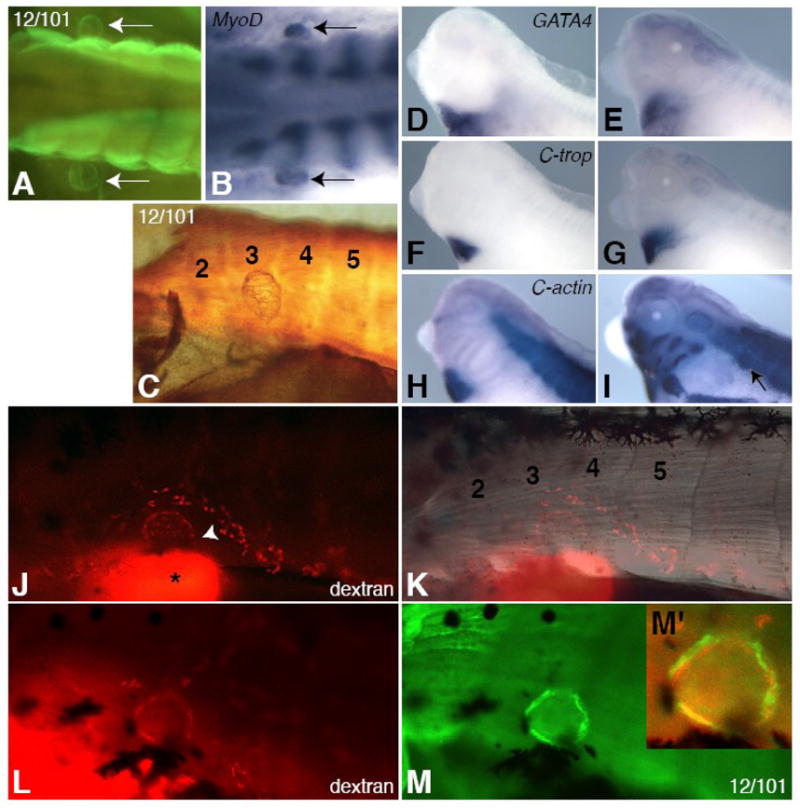
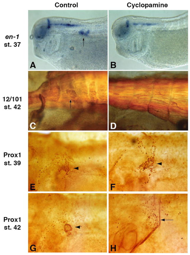
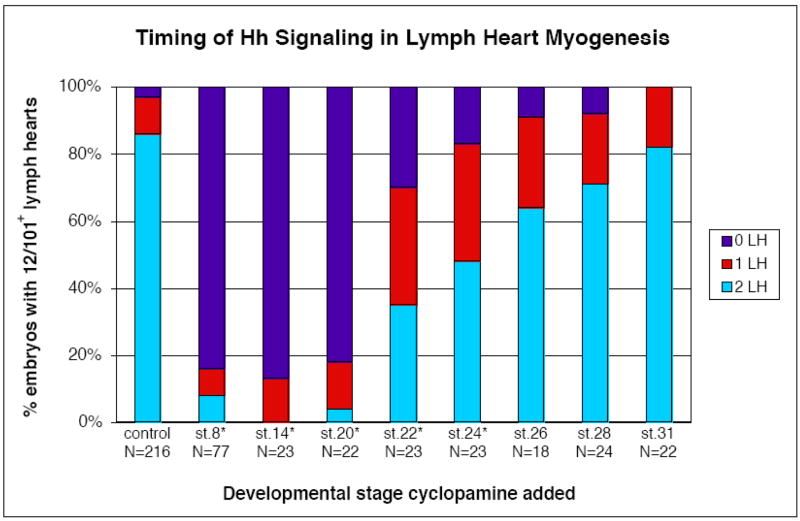
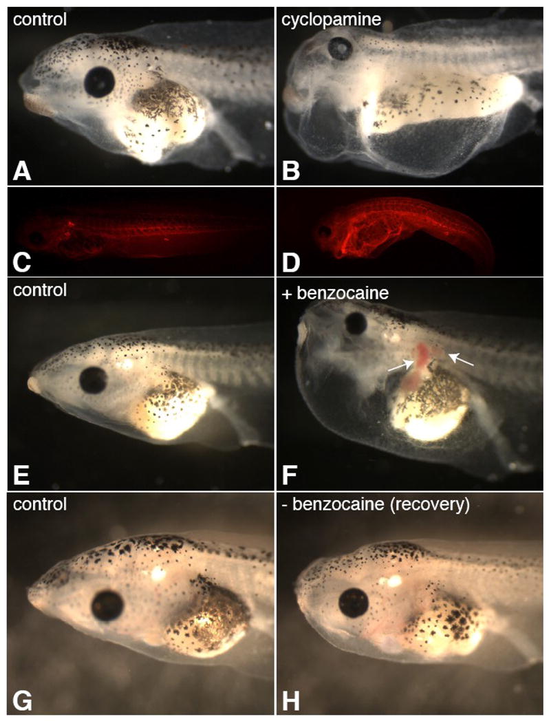
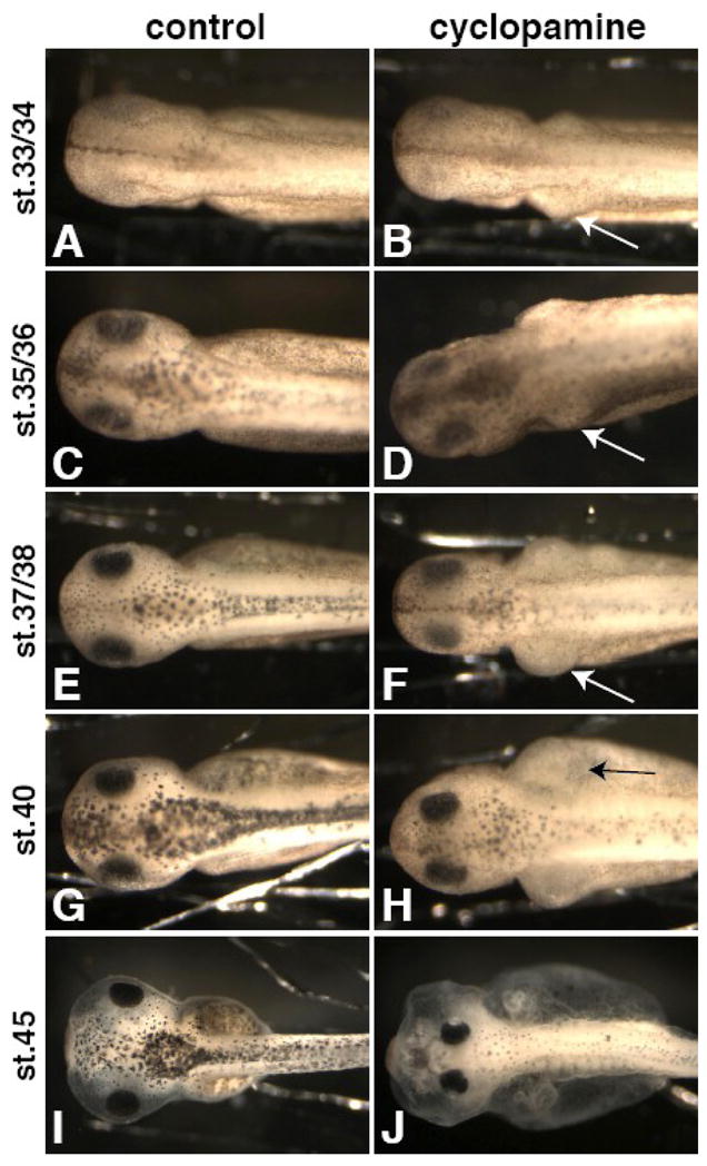
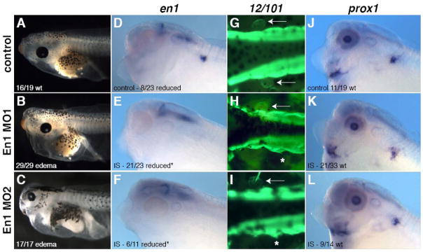
Lymph hearts are pulsatile organs, present in lower vertebrates, that function to propel lymph into the venous system. Although they are absent in mammals, the initial veno-lymphatic plexus that forms during mammalian jugular lymph sac development has been described as the vestigial homologue of the nascent stage of ancestral anterior lymph hearts. Despite the widespread presence of lymph hearts among vertebrate species and their unique function, extremely little is known about lymph heart development. We show that Xenopus anterior lymph heart muscle expresses skeletal muscle markers such as myoD and 12/101, rather than cardiac markers. The onset of lymph heart myoblast induction can be visualized by engrailed-1 (en1) staining in anterior trunk somites, which is dependent on Hedgehog (Hh) signaling. In the absence of Hh signaling and upon en1 knockdown, lymph heart muscle fails to develop, despite the normal development of the lymphatic endothelium of the lymph heart, and embryos develop edema. These results suggest a mechanism for the evolutionary transition from anterior lymph hearts to jugular lymph sacs in mammals.


Figures

Figure 1
Figure 1. Lymph heart myoblasts express skeletal muscle markers and not cardiac markers
The 12/101 antibody marks lymph hearts in stage 42 tadpoles (A, C, M) and MyoD is expressed in differentiating lymph hearts at stage 40 (B, arrows). Two bilaterally symmetrical lymph hearts can be seen adjacent to the somites (A, B, arrows). The lymph heart forms in a region adjacent to somites 3 and 4 (C, K, trunk somites are numbered). Lymph heart muscle consists of a meshwork of myotubes (C). The lymph heart does not express cardiac markers GATA4 (D, E), cardiac troponin (F, G), or cardiac actin (H, I) at st. 33/34 (D, F, H) or st. 40 (E, G, I), except for cardiac actin at st. 40 (I, arrow), which marks all immature muscle (note expression in paraxial and head mesoderm in H, I). Rhodamine-dextran (mini-Ruby) injected subcutaneously into the posterior ventral tail fin drains anteriorly into the kidney (J, bright spot with asterisk) and into the lymph heart through vessels (J, arrowhead). The labeled lymph heart is located between somites 3 and 4 (K). Embryos injected with rhodamine-dextran (L, red) and then stained for the skeletal muscle antibody 12/101 (M, green) show co-expression (M′, yellow). All lateral views, anterior left, except for (A, B), which are dorsal views.
Figure 2
Figure 2. en1 and prox1 label the developing lymph heart musculature and endothelial tissue, respectively
The expression of en1 is localized to the mid-hindbrain boundary, spinal interneurons, and anterior somites. The onset of somitic expression occurs at stage 28 (A) in a superficial region on a horizontal plane with the notochord (E, arrowhead, middle of notochord indicated by red line). The expression does not overlap with 12/101 staining in the differentiated muscle (green). Lateral views show early expression in the anterior somites (A–B, arrowheads), which intensifies and moves ventrally to occupy the final position of the lymph heart (C–D, G). A transverse section of a stage 33 tadpole (F) shows the intensity of expression increasing. By stage 37 (G), en1-positive cells have moved ventrally relative to the position of the notochord (red line) and are found directly above the glomus (arrow) and pronephric tubules (arrowhead). At stage 40, prox1 RNA (blue, H, J, L) and Prox1 protein (brown, I, K, M) are localized to the developing endothelial tissue of the lymph heart (H, I, arrows). (J, K) Higher magnification views of H and I illustrate the budding lymphatic vasculature dorsal to the lymph heart (arrowheads). (L) The expression of prox1 is slightly ventral to the notochord (red line) and dorsal to the pronephroi (arrow) and does not overlap with differentiated muscle (12/101, green). (M) Prox1 (brown) and en1 (blue) expressing cells co-localize at the site of lymph heart formation (arrow). The epidermis has sloughed off during the processing of this embryo.
Figure 3
Figure 3. Lymph heart muscle but not lymphatic endothelium requires Hh signaling
Cyclopamine treatment greatly reduces en1 expression in the somites (23 out of 24 embryos), while neural expression is relatively unaffected (B compared to A, arrow in A indicates lymph heart expression). At later stages, 12/101 staining shows that lymph heart musculature is missing in cyclopamine-treated tadpoles (D compared to C, arrow in C indicated lymph heart muscle). The presence of Prox1-positive lymphatic endothelial cells (arrowheads) is not affected by cyclopamine treatment at stage 39 (F compared to E) or stage 42 (H compared to G). (H) The loss of lymph heart musculature leads to a less compact distribution of Prox1-positive cells in the lymph heart (arrow).
Figure 4
Figure 4. The timing of Hedgehog induction of lymph hearts corresponds to the onset of en1 expression
Cyclopamine or ethanol (as a solvent control) was added to embryos at the specified developmental stages; tadpoles were fixed at stage 42–45, and stained with 12/101 for presence of lymph heart musculature. The ethanol controls for all stages are combined in this graph, but χ2 tests were performed between stage-matched cyclopamine and control samples for determination of statistical significance. Cyclopamine treatment has a less potent effect when added after stage 20 (4% wildtype at st.20 vs. 35% wildtype at st.22). Experimental and control samples are not significantly different when embryos are added to cyclopamine at stage 26, just prior to en1 expression in lymph heart myoblasts, or later in development (*p<0.01).
Figure 5
Figure 5. Lymph heart paralysis results in reversible edema
Cylopamine treatment from stage 8 results in loss of lymph heart beating and severe edema at stage 42 (B; 100%, N=79). Florescent dye injected subcutaneously into the fin of cyclopamine-treated tadpoles (stage 42) fills the body cavity and fails to clear the body in an hour (D), unlike in controls (C). Tadpoles with visibly beating lymph hearts (st.42+) were placed in a dilute benzocaine solution (0.005–0.01%), which resulted in lymph heart paralysis while leaving the cardiac heartbeat unaffected. After 24 hours of lymph heart paralysis, tadpoles developed severe edema with blood pooling in the kidney, lymph heart, and gills (F, arrows; 100%, N=60), while control tadpoles did not (E). Removal of these embryos from benzocaine restored lymph heart function and tadpoles were mostly recovered in 4 days (H; 58%, N=57, compare to G, control).
Figure 6
Figure 6. The development of edema upon cyclopamine treatment does not correspond to lymph heart beating
Embryos were added to cyclopamine (B, D, F, H, J) at stage 8 and compared to controls (A, C, E, G, I) at stage 33/34 (A, B), st. 35/36 (C, D), st. 37/38 (E, F), st. 40 (G, H), and st. 45 (I, J). Edema begins around the kidney area as early as st. 33/34 (B, arrow) and continues to worsen (D, F, H, arrows), long before the lymph hearts start beating (st. 41–42).
Figure 7
Figure 7. Engrailed-1 is required for lymph heart myogenesis
Two non-overlapping MOs were designed to block en1 translation. Bilateral injection of En1 MO leads to loss of lymph heart beat and edema (A–C). Unilateral injection of En1 MO leads to specific loss of lymph heart musculature assayed by en1 expression (D–F, *p<0.05 χ2 test) and 12/101 staining (G–I, dorsal view, arrow points to lymph heart, asterisk indicates injected side). In controls, 100% of embryos have two 12/101-positive lymph hearts (N=43), while 5% of En1 morphants do (En1 MO1 N=42, En1 MO2 N=38). Lymph heart endothelium (prox1, J–L) is not affected by En1 knockdown compared to controls (p>0.05, χ2 test).

Similar articles

Cited by

References

    1. Cheng L, Alvares LE, Ahmed MU, El-Hanfy AS, Dietrich S. The epaxial-hypaxial subdivision of the avian somite. Dev Biol. 2004;274:348–69. - PubMed
    1. Cleaver O, Tonissen KF, Saha MS, Krieg PA. Neovascularization of the Xenopus embryo. Dev Dyn. 1997;210:66–77. - PubMed
    1. Currie PD, Ingham PW. Induction of a specific muscle cell type by a hedgehog-like protein in zebrafish. Nature. 1996;382:452–5. - PubMed
    1. Davis CA, Holmyard DP, Millen KJ, Joyner AL. Examining pattern formation in mouse, chicken and frog embryos with an En-specific antiserum. Development. 1991;111:287–98. - PubMed
    1. Devic E, Paquereau L, Vernier P, Knibiehler B, Audigier Y. Expression of a new G protein-coupled receptor X-msr is associated with an endothelial lineage in Xenopus laevis. Mech Dev. 1996;59:129–40. - PubMed

Publication types

MeSH terms

LinkOut - more resources