. 2010 Mar 15;187(1):129-37.
doi: 10.1016/j.jneumeth.2010.01.002. Epub 2010 Jan 11.

Regional convection-enhanced delivery of gadolinium-labeled albumin in the rat hippocampus in vivo

Affiliations

Regional convection-enhanced delivery of gadolinium-labeled albumin in the rat hippocampus in vivo

Garrett W Astary et al. J Neurosci Methods. .

Abstract

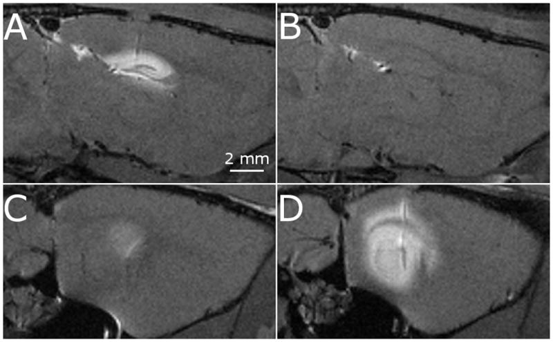
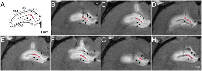
Convection-enhanced delivery (CED) has emerged as a promising method of targeted drug delivery for treating central nervous system (CNS) disorders, but the influence of brain structure on infusate distribution is unclear. We have utilized this approach to study extracellular transport and distribution of a contrast agent in the hippocampus, a complex structure susceptible to CNS disorders. The magnetic resonance (MR) contrast agent diethylene triamene penta-acetic acid chelated gadolinium-labeled albumin (Gd-albumin), tagged with Evans blue dye, was directly infused (V(i)=5 microl) into the dorsal and ventral hippocampus of seven male Sprague-Dawley rats. The final distribution profile of the contrast agent, a product of CED and limited diffusion, was observed in vivo using high-resolution T1-weighted MR imaging at 11.1T. Dense cell layers, such as the granule cell layer of the dentate gyrus and the pyramidal cell layer of CA1, appeared to be barriers to transport of the tracer. Three-dimensional distribution shape and volume (V(d)) differences, between the dorsal and ventral hippocampus infusions, were determined from the MR images using a semi-automatic segmentation routine (dorsal V(d)=23.4+/-1.8 microl, ventral V(d)=36.4+/-5.1 microl). Finer structural detail of the hippocampus was obtained using a combination of histological analysis and fluorescence imaging. This study demonstrates that CED has the potential to target all regions of the hippocampus and that tracer distribution is influenced by infusion site, underlying structure and circuitry, and extent of backflow. Therefore, CED, combined with high-resolution MR imaging, may be a useful strategy for delivering therapeutics for the treatment of CNS disorders affecting the hippocampus.


Figures

Fig. 1
Fig. 1
Damage induced by the infusion cannula in the dorsal hippocampus. (A) Blood at the tip of the cannula, (B) Blood within the alveus/corpus-collosum boundary
Fig. 2
Fig. 2
Sagittal images of a single rat demonstrating the apparent disconnect between the dorsal hippocampus (top row) and ventral hippocampus (bottom row). The disconnect is seen when the contrast agent is infused into the dorsal hippocampus (A and C) and ventral hippocampus (B and D).
Fig. 3
Fig. 3
High-resolution T1-weighted MR images of dorsal hippocampus infusions. (A) Schematic of key structures in the dorsal hippocampus adapted from (Paxinos and Watson, 1998). (B-H) MR image coronal slice of infusion site for dorsal hippocampus infusions in 7 rats. Filled arrow heads, dentate gyrus granule cell layer; unfilled arrow heads, CA1 pyramidal cell layer; asterisk, hippocampal fissure.
Fig. 4
Fig. 4
High-resolution T1-weighted MR images of ventral hippocampus infusions. (A) Schematic of key structures in the ventral hippocampus adapted from (Paxinos and Watson, 1998). (B-H) MR image coronal slice of ventral hippocampus infusions into 7 rats. Filled arrow heads, dentate gyrus granule cell layer; unfilled arrow heads, CA1 pyramidal cell layer; asterisk, hippocampal fissure, filled arrow, alveus; unfilled arrow, corpus collosum.
Fig. 5
Fig. 5
High-resolution T1-weighted MR image coronal slice of the dorsal hippocampus distribution volume (left image in subfigure) and ventral hippocampus (right image in subfigure) and corresponding segmentation (middle image in subfigure) for 4 rats (A-D).
Fig. 6
Fig. 6
Histological images following infusate CED into the dorsal hippocampus showing Evans blue dye spreading throughout the dorsal hippocampus. (A, C) Fluorescence images of 2 subjects showing limited penetration in the dorsal hippocampal dense granule cell layer (filled arrowhead) and pyramidal cell layer (unfilled arrowhead). Preferential distribution can be seen in the hippocampal fissure (asterisks) and alveus (filled arrow). (B) Black-gold stained image in close proximity to (A) confirming alveus and dense cell layer approximations. (D) Cresyl violet staining of a section in close proximity to (C) confirming dense cell layers.
Fig. 7
Fig. 7
Histological images following infusate CED into the ventral hippocampus showing Evans blue dye spreading thoughout the ventral hippocampus. Arrowheads denote granule cell layer of the dentate gyrus and pyramidal cell layer of the CA1. (A) A fluorescence image of Evans blue seen preferentially in the ventral hippocampal fissure (asterisks), alveus (filled arrow) and corpus collosum (unfilled arrow). (B) Cresyl violet stained image of a section in close proximity to (A). (C) Fluorescent image of Evans blue seen preferentially in the alveus and corpus collosum. Chevron shows Evans blue in the perivascular space. (D) Cresyl violet stained image of a section in close proximity to (C).

Similar articles

Cited by

References

    1. Abujudeh HH, Kaewlai R, Kagan A, Chibnik LB, Nazarian RM, High WA, Kay J. Nephrogenic systemic fibrosis after gadopentetate dimeglumine exposure: case series of 36 patients. Radiology. 2009;253:81–9. - PubMed
    1. Andersen AH. On the Rician distribution of noisy MRI data. Magn Reson Med. 1996;36:331–3. - PubMed
    1. Andersen P, Bliss TV, Lomo T, Olsen LI, Skrede KK. Lamellar organization of hippocampal excitatory pathways. Acta Physiol Scand. 1969;76:4A–5A. - PubMed
    1. Auer RN, Siesjo BK. Biological differences between ischemia, hypoglycemia, and epilepsy. Ann Neurol. 1988;24:699–707. - PubMed
    1. Bertram EH. Temporal lobe epilepsy: where do the seizures really begin? Epilepsy Behav. 2009;14 (Suppl 1):32–7. - PMC - PubMed

Publication types

MeSH terms

LinkOut - more resources