. 2010 Apr 1;19(7):1230-47.
doi: 10.1093/hmg/ddq001. Epub 2010 Jan 12.

A mutation in the mouse Amelx tri-tyrosyl domain results in impaired secretion of amelogenin and phenocopies human X-linked amelogenesis imperfecta

Affiliations

A mutation in the mouse Amelx tri-tyrosyl domain results in impaired secretion of amelogenin and phenocopies human X-linked amelogenesis imperfecta

Martin J Barron et al. Hum Mol Genet. .

Abstract

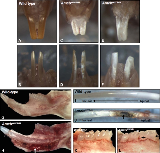
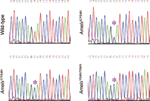
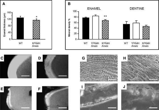
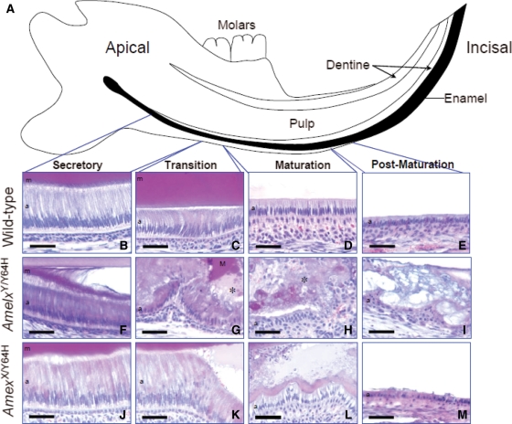
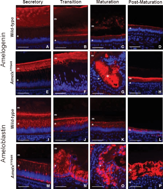
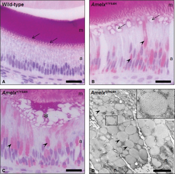
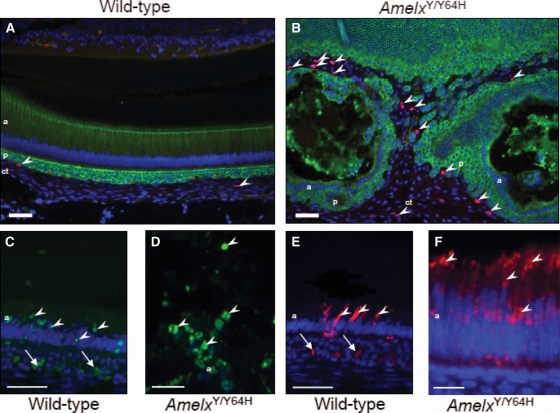
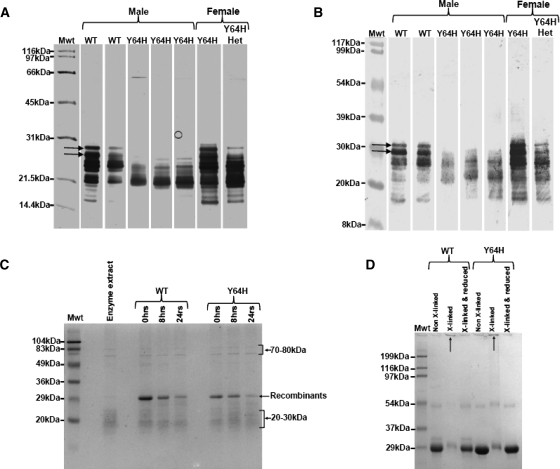
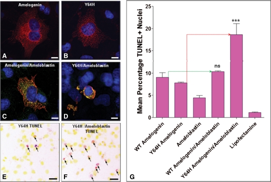
Amelogenesis imperfecta (AI) describes a broad group of clinically and genetically heterogeneous inherited defects of dental enamel bio-mineralization. Despite identification of a number of genetic mutations underlying AI, the precise causal mechanisms have yet to be determined. Using a multi-disciplinary approach, we describe here a mis-sense mutation in the mouse Amelx gene resulting in a Y --> H substitution in the tri-tyrosyl domain of the enamel extracellular matrix protein amelogenin. The enamel in affected animals phenocopies human X-linked AI where similar mutations have been reported. Animals affected by the mutation have severe defects of enamel bio-mineralization associated with absence of full-length amelogenin protein in the developing enamel matrix, loss of ameloblast phenotype, increased ameloblast apoptosis and formation of multi-cellular masses. We present evidence to demonstrate that affected ameloblasts express but fail to secrete full-length amelogenin leading to engorgement of the endoplasmic reticulum/Golgi apparatus. Immunohistochemical analysis revealed accumulations of both amelogenin and ameloblastin in affected cells. Co-transfection of Ambn and mutant Amelx in a eukaryotic cell line also revealed intracellular abnormalities and increased cytotoxicity compared with cells singly transfected with wild-type Amelx, mutant Amelx or Ambn or co-transfected with both wild-type Amelx and Ambn. We hypothesize that intracellular protein-protein interactions mediated via the amelogenin tri-tyrosyl motif are a key mechanistic factor underpinning the molecular pathogenesis in this example of AI. This study therefore successfully links phenotype with underlying genetic lesion in a relevant murine model for human AI.


Figures

Figure 1.
Figure 1.
Morphology of the dentition of Amelx mutant mice. In wild-type (WT) maxillary (A) and mandibular (B) incisor teeth, the enamel appeared smooth, opalescent and orange/brown in colour. (C and D) In heterozygous female (AmelxX/Y64H) mutant mice, the incisor teeth showed patchy regions of roughened, chalky white enamel. (E and F) In hemizygous male (AmelxY/Y64H) mutant mice, the entire enamel surface of the incisor teeth was roughened, opaque and chalky white. Frequently, the incisor teeth of affected mice were shortened with irregular incisal edges (C–F). (G) The inferior border of the WT mandible appeared smooth and unremarkable; (H) in contrast, that of AmelxY/Y64H mutant mice was enlarged, eroded and discoloured containing a large mass of soft tissue (arrow). (I) The surface of dissected WT mandibular incisors was smooth with progressive maturation from white, incompletely mineralized enamel at the apex of the tooth to increasingly pigmented, fully mineralized enamel towards the incisal tip. (J) The surface of mandibular incisors dissected from AmelxY/Y64H mice, at the point where enamel maturation would normally occur, was irregular and discoloured (arrow) with ridges running along the long axis of the tooth. (K) The molar teeth of WT mice were smooth and opalescent whereas (L) those of AmelxY/Y64H mice were opaque and roughened (arrows) with abrasion of the cusps (asterisk).
Figure 2.
Figure 2.
Identification of the Amelx Y64H mutation. Partial sequence chromatograms of Amelx depicting the WT sequence, and the T → C transition (asterisks) that leads to the Y64H mutation in amelogenin in heterozygous female (AmelxX/Y64H), hemizygous male (AmelxY/Y64H) and homozygous female (AmelxY64C/Y64H) mutant mice.
Figure 3.
Figure 3.
Enamel phenotype of Amelx mutant mice. (A) Enamel thickness in heterozygous female (AmelxX/Y64H) mutant mice was significantly reduced compared with WT (P < 0.05). Enamel thickness in hemizygous males could not be reliably measured due to large areas of enamel being missing from the teeth and thus no data are presented. (B) Enamel mineral density in AmelxX/Y64H females was also significantly reduced compared with WT littermates (P < 0.001). Mineral density in hemizygous males was, however, not significantly different from WT. There were no significant differences in mineral levels in the dentine of the teeth of affected animals compared with WT tissue. (CF) Representative transverse sections of mature enamel prepared for quantitative microradiography clearly show qualitative differences in enamel from WT (C), heterozygous females (AmelxX/Y64H) (D) and hemizygous males (AmelxY/Y64H) (E, F). Scale bars = 100 µm. (GJ) Scanning electron micrographs of WT enamel (G) exhibited decussating prism architecture characteristic of rodent enamel. Enamel in AmelxX/Y64H females also showed decussating prisms (H) but they were less highly ordered and possessed a more open structure compared with WT. Enamel from AmelxY/Y64H affected males could only be imaged where remnants of the tissue were present and revealed severely dysplastic non-prismatic ‘enamel’ with a smooth glass-like appearance (I, J). Scale bars = 20 µm.
Figure 4.
Figure 4.
Histological analysis of the lower incisor teeth. (A) Diagrammatic representation of the mouse lower incisor tooth within the mandible. As indicated in black, enamel production commences at the apical region of the tooth and proceeds towards the incisal edge. (BE) The secretory, transition, maturation and post-maturation stages of amelogenesis showed characteristically normal morphology in the WT lower incisor tooth. (F) The secretory ameloblasts of hemizygous male (AmelxY/Y64H) mutant mice differentiated normally but soon lost contact with the newly secreted enamel matrix and became abnormally eosinophilic. (G and H) In the transition and maturation zones, the ameloblasts of AmelxY/Y64H mice adopted a highly abnormal morphology and formed irregular, multi-cellular masses. In these regions, large quantities of amorphous material containing cellular debris (asterisks) were apparent. (I) The multi-cellular masses frequently persisted through the post-maturation zone to the gingival margin. (J) Although the secretory ameloblasts of heterozygous female (AmelxX/Y64H) mutant mice appeared largely normal, a proportion exhibited increased eosinophilia. (K and L) In the transition and maturation zones, contact of the ameloblasts with the enamel matrix was occasionally lost leading to the formation of blister-like structures. (M) In contrast, the appearance of the post-maturation ameloblasts resembled that of WT mice. m, enamel matrix; a, ameloblasts. Scale bars, 50 µm.
Figure 5.
Figure 5.
Amelogenin and ameloblastin distribution in Amelx mutant mice. (AH) Amelogenin distribution. (A) Strong amelogenin immunolabelling of the enamel matrix, arranged as closely packed parallel lines, was observed in the secretory zone of WT mice. (B) Enamel matrix degradation commences in the transition zone and this was reflected in the WT enamel matrix where the intensity of immunostaining was markedly reduced. (C) Little amelogenin immunoreactivity was detected in the maturation zone where the process of enamel matrix degradation is completed. (D) No amelogenin immunoreactivity was observed in the post-maturation enamel matrix. (E) In the secretory zone of hemizygous male (AmelxY/Y64H) mutant mice strong amelogenin immunolabelling of the enamel matrix was observed; however, the pattern of parallel lines observed in WT mice was lost. Additionally, the secretory ameloblasts of AmelxY/Y64H mice showed increased immunostaining in the cytoplasm. (F and G) Where the ameloblasts became disorganized in AmelxY/Y64H mice, strong amelogenin immunostaining was detected both in the enamel matrix and within the multi-cellular masses (asterisk). (H) Amelogenin immunoreactivity was often detected in the post-maturation zone where enamel matrix is normally absent. (IP) Ameloblastin distribution. (I) In the secretory zone of WT mice, strong ameloblastin immunostaining of the enamel matrix was detected in the vicinity of the ameloblast Tomes’ processes. Strong ameloblastin immunoreactivity was also observed in the supranuclear compartment of the secretory ameloblasts. (J–L) Ameloblastin expression in the cytoplasm of WT ameloblasts was detected throughout the subsequent stages of amelogenesis. (M) In hemizygous male (AmelxY/Y64H) mutant mice, the secretory ameloblasts were immunoreactive for ameloblastin; however, the regular pattern of supra-nuclear immunoreactivity observed in WT mice was replaced by patchy aggregates of ameloblastin immunostaining. Immunolabelling of the enamel matrix was very weak in AmelxY/Y64H mice. (N and O) Where the ameloblasts became disorganized in those regions corresponding to the transition and maturation zones of WT mice, intracellular ameloblastin immunostaining became more intense in AmelxY/Y64H mice. (P) Extracellular ameloblastin immunoreactivity was frequently observed in the post-maturation stage of enamel formation in AmelxY/Y64H mice. m, enamel matrix; a, ameloblasts. Scale bars, 50 µm.
Figure 6.
Figure 6.
Acrylic resin histology and immunoelectron microscopy. (A) Semi-thin, transverse sections of the secretory zone of WT mice revealed pale-staining ameloblasts and an intensely eosinophilic, enamel matrix. The interdigitations of the ameloblast Tomes’ processes were clearly evident in the region where new enamel matrix was forming (arrows). (B and C) In hemizygous male (AmelxY/Y64H) mutant mice, many of the ameloblasts contained large numbers of eosinophilic vesicles (arrowheads). Although the enamel matrix was intensely eosinophilic as observed in WT mice, it was highly disorganized (asterisk) and contact with the ameloblasts was lost. (C) Where new enamel matrix was being secreted, the interdigitating pattern seen in WT mice was replaced by large vacuolar structures (arrows). (D) Immunoelectron microscopy of AmelxY/Y64H secretory ameloblasts revealed the cytoplasm to be engorged by vacuoles of up to 2 µm diameter which were present in such large numbers that they led to the ameloblast nuclei becoming distorted. These vacuoles were strongly immunoreactive (black dots) for amelogenin (inset). m, enamel matrix; a, ameloblasts; n, nuclei. Scale bars in A–C, 25 µm and in D, 2 µm.
Figure 7.
Figure 7.
Analysis of cell proliferation and apoptosis. (A) Pan-cytokeratin (green) and BrdU (red) dual immunolabelling of the enamel organ of WT mice showing occasional BrdU-positive nuclei (arrowheads), confined to the underlying connective tissue compartment. (B) In the multi-cellular masses observed in hemizygous male (AmelxY/Y64H) mutant mice, BrdU-positive nuclei remained confined to the connective tissue element (arrowheads). (C) TUNEL-positive nuclei (arrowheads) were observed in the transition zone of WT mice. (D) In hemizygous male (AmelxY/Y64H) mutant mice, numerous TUNEL-positive nuclei (arrowheads) were observed in the amorphous material associated with the multi-cellular masses. (E) Immunolabelling for activated caspase 3 in WT mice revealed strong immunoreactivity in transition stage ameloblasts (arrowheads). (F) The ameloblasts immediately proximal to the area where multi-cellular masses form in AmelxY/Y64H mice were strongly immunoreactive for activated caspase 3 (arrowheads). The arrows in (C) and (E) indicate autofluorescence from erythrocytes. a, ameloblasts; p, stratum intermedium/papillary layer complex; ct, connective tissue. Scale bars in A–C and E, 50 µm and in D and F, 25 µm.
Figure 8.
Figure 8.
Biochemical investigations relating to the Y64H developing enamel proteome. (A) SDS–PAGE of secretory stage developing enamel removed from WT and Y64H mouse mandibular incisors showing apparent lack of full-length amelogenin proteins in the mutant animals (arrowed). Male and female WT animals had clear evidence of higher molecular weight amelogenins migrating up to ∼30 kDa. Y64H affected males had no proteins corresponding to this molecular weight range. Female mice, heterozygous with respect to the Y64H mutation did have higher molecular weight amelogenin present but at reduced amounts compared with WT. (B) Western blot of secretory stage developing enamel removed from WT and Y64H mouse mandibular incisors confirming the apparent lack of full-length amelogenin proteins (arrowed) in the mutant animals observed by SDS–PAGE (A). (C) SDS–PAGE of WT and Y64H recombinant amelogenins before and after incubation with secretory enamel protease extracts. After 24 h, Coomassie Blue staining revealed clear evidence of proteolytic degradation for both recombinant proteins, with progressive loss of intact protein (SDS Mr = 29 kDa). Proteins migrating at 70–80 kDa and around 20 kDa were also present in the MMP20 extracts and can be ignored with regard to the degradation of the recombinants. (D) SDS–PAGE of WT and Y64H recombinant amelogenins subjected to near-neighbour molecular cross-linking. Both WT and Y64H recombinant amelogenins were cross-linked to form supra-molecular complexes that failed to penetrate the SDS–PAGE stacking gel. On reduction of the cross-links, the amelogenins forming these complexes were returned to their monomeric state migrating at around 29 kDa.
Figure 9.
Figure 9.
Effect of the Y64H mutant amelogenin on cell survival. (A) COS-7 cell transfected with the WT amelogenin construct. The cell shows strong amelogenin expression (red fluorescence) and appears large and viable. (B) A similar cell transfected with Y64H amelogenin construct showing strong amelogenin expression (red fluorescence) is also typically large and viable. (C) A cell co-transfected with WT amelogenin and ameloblastin constructs shows strong immunolabelling for both proteins (orange–yellow fluorescence) and also appears large and viable. (D) Cells co-transfected with Y64H mutant amelogenin and ameloblastin constructs (orange–yellow fluorescence) were typically small with a condensed nucleus (blue) consistent with apoptosis. (E and F) TUNEL analysis of cells transfected with (E), mutant amelogenin alone and (F), mutant amelogenin and ameloblastin constructs. To improve discrimination of apoptotic cell nuclei, the images of the cells subjected to TUNEL analysis were pseudo-coloured such that TUNEL-positive nuclei appear red (arrows) and normal nuclei appear yellow. Scale bars represent 10 µm. (G) Results of TUNEL assays on four replicate experiments of each transfection/co-transfection. One-way ANOVA followed by a Bonferroni multiple comparison test showed that the large increase in mean percentage of TUNEL-positive nuclei detected in the Y64H mutant amelogenin/ameloblastin co-transfections was statistically significant (P < 0.0001) compared with cells transfected with Y64H mutant amelogenin alone. No statistically significant difference in the incidence of apoptosis could be shown between cells transfected with WT amelogenin alone and those co-transfected with WT amelogenin and ameloblastin.

Similar articles

Cited by

References

    1. Backman B., Holm A.K. Amelogenesis imperfecta: prevalence and incidence in a northern Swedish county. CommunITY Dent. Oral Epidemiol. 1986;14:43–47. - PubMed
    1. Backman B. Amelogenesis imperfecta—clinical manifestations in 51 families in a northern Swedish county. Scand. J. Dent. Res. 1988;96:505–516. - PubMed
    1. Witkop C.J., Jr Amelogenesis imperfecta, dentinogenesis imperfecta and dentin dysplasia revisited: problems in classification. J. Oral Pathol. 1988;17:547–553. - PubMed
    1. Stephanopoulos G., Garefalaki M.E., Lyroudia K. Genes and related proteins involved in amelogenesis imperfecta. J. Dent. Res. 2005;84:1117–1126. - PubMed
    1. Hu J.C., Chun Y.H., Al Hazzazzi T., Simmer J.P. Enamel formation and amelogenesis imperfecta. Cells Tissues Organs. 2007;186:78–85. - PubMed

Publication types

MeSH terms