. 2010 Mar;19(3):288-99.
doi: 10.1177/0961203309353773. Epub 2010 Jan 12.

Lupus-prone New Zealand Black/New Zealand White F1 mice display endothelial dysfunction and abnormal phenotype and function of endothelial progenitor cells

Affiliations

Lupus-prone New Zealand Black/New Zealand White F1 mice display endothelial dysfunction and abnormal phenotype and function of endothelial progenitor cells

S G Thacker et al. Lupus. 2010 Mar.

Abstract

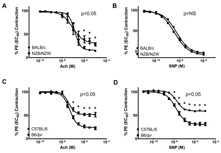
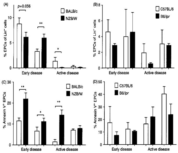
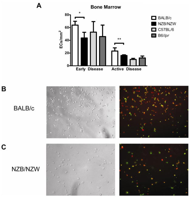
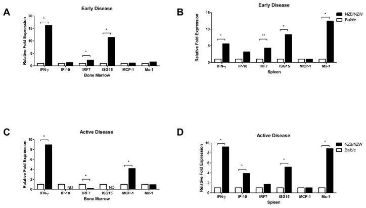
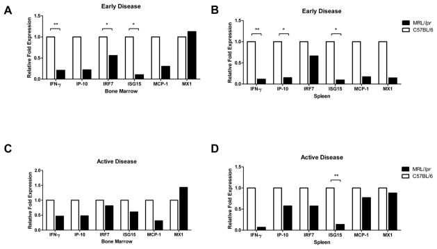
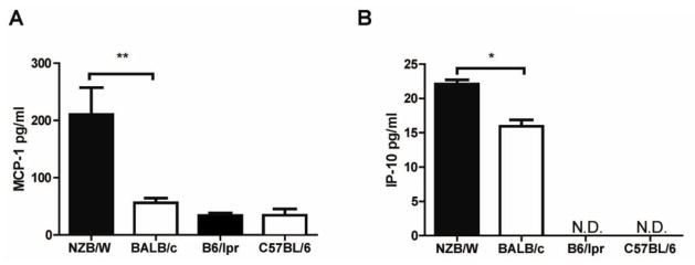
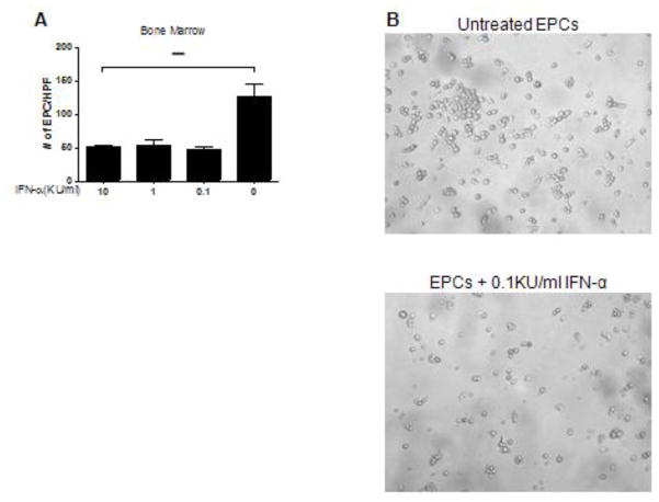
Patients with systemic lupus erythematosus (SLE) have an impairment in phenotype and function of endothelial progenitor cells (EPCs) which is mediated by interferon alpha (IFN-alpha). We assessed whether murine lupus models also exhibit vasculogenesis abnormalities and their potential association with endothelial dysfunction. Phenotype and function of EPCs and type I IFN gene signatures in EPC compartments were assessed in female New Zealand Black/New Zealand White F(1) (NZB/W), B6.MRL-Fas(lpr)/J (B6/lpr) and control mice. Thoracic aorta endothelial and smooth muscle function were measured in response to acetylcholine or sodium nitropruside, respectively. NZB/W mice displayed reduced numbers, increased apoptosis and impaired function of EPCs. These abnormalities correlated with significant decreases in endothelium-dependent vasomotor responses and with increased type I IFN signatures in EPC compartments. In contrast, B6/lpr mice showed improvement in endothelium-dependent and endothelial-independent responses, no abnormalities in EPC phenotype or function and downregulation of type I IFN signatures in EPC compartments. These results indicate that NZB/W mice represent a good model to study the mechanisms leading to endothelial dysfunction and abnormal vasculogenesis in lupus. These results further support the hypothesis that type I IFNs may play an important role in premature vascular damage and, potentially, atherosclerosis development in SLE.


Conflict of interest statement

Competing interests

The author(s) declare that they have no competing interests

Figures

Figure 1
Figure 1. Decreased endothelial-mediated vasorelaxation in NZB/W mice
Results assess acetylcholine (Ach)-mediated endothelium-dependent relaxation and sodium nitroprusside (SNP)-mediated endothelium-independent relaxation in aortic rings from (A), (B) 36-week-old NZB/W and BALB/c mice and (C), (D) 16-week-old B6/lpr and C57BL/6 mice. NZB/W mice have impaired Ach-mediated responses, while B6/lpr mice showed enhanced ACh-mediated and SNP-mediated responses. Results are mean ± SEM % phenylephrine (PE) EC80 contraction (n= 5–8 mice/group). *p<0.05; NS=not significant.
Figure 2
Figure 2. EPCs are decreased in NZB/W mice and show higher levels of apoptosis
EPCs were quantified in the bone marrow and spleen in pre-nephritic (early disease) and nephritic (active disease) NZB/W (A) and B6/lpr mice (B), as well as control mice. A significantly higher number of NZB/W EPCs were apoptotic, as determined by Annexin V expression at (C) pre-nephritic (early) time-point, both in the bone marrow and the spleen and at the nephritic (late) time point in the bone marrow. The B6/lpr mice showed no significant increases in EPC apoptosis at D) the early or active disease time points. Results are mean ± SEM % lineage-negative EPCs (n= 5–8 mice/group). *p<0.05; **p<0.01.
Figure 3
Figure 3. NZB/W EPCs exhibit impaired capacity to differentiate into mature ECs
Bone marrow-derived EPCs were cultured under proangiogenic conditions and incubated at different time-points during culture with Dil-ac-LDL and BS-1-FITC. Mature endothelial cells were identified by co-staining of BS-1 and ac-LDL. (A) Bar graphs represent the number of mature endothelial cells per centimeter at day 7, when comparing NZB/W and B6/lpr bone marrow-derived EPCs with control EPCs, at early and active disease time-points. Results are mean ± SEM of 3 or 4 independent experiments; *p<0.05, **p<0.01. (B), (C) Results are representative images obtained from BALB/c (B) and NZB/W (C) EPCs cultured under proangiogenic conditions for 7 days. Left panels show bright field images and right panels show images obtained by fluorescent microscopy. NZB/W EPCs show decreased ability to differentiate into endothelial cells, when compared to BALB/c mice. DiI-Ac-LDL is red while BS-1 lectin is green. Total magnification is ×100.
Figure 4
Figure 4. Type I IFN-inducible genes are increased in NZB/W EPC compartments
Results are displayed as relative fold change of type I IFN-inducible genes over control BALB/c mice (n=5 mice/group) in bone marrow and spleen EPC compartments during (A), (B) early and (C), (D) active disease stage. Transcripts were normalized to β-actin; *p<0.05; **p<0.01; ND = not detected.
Figure 5
Figure 5. B6/lpr EPCs display decreased expression of type I IFN-inducible genes
Results are displayed as relative fold change of type I IFN-inducible genes over control C57BL/6 mice (n=3 mice/group) in bone marrow and spleen EPC compartments during (A), (B) early and (C), (D) active disease stage. Transcripts were normalized to β-actin; *p<0.05; **p<0.01.
Figure 6
Figure 6. Increased circulating levels of type I-IFN inducible proteins in NZB/W mice
Levels of the type I IFN-sensitive chemokines MCP-1 and IP-10 were quantified in the plasma of lupus-prone mice with active disease. Results represent mean ± SEM (n = 3–4 mice per group). *p<0.05.
Figure 7
Figure 7. IFN-α is toxic to murine EPCs
Bone marrow EPCs from BALB/c mice were cultured in proangiogenic media in the presence or absence of graded concentrations of recombinant IFN-α for 3 days. Numbers of mature ECs were quantified at 7 days in culture. Exposure to IFN-α resulted in a significant loss of EPC ability to differentiate into mature endothelial cells. (A) Bar graph displays 1 representative experiment and represent mean ± SEM of 5 high-power fields; n=3, ***p<0.001. (B) Representative bright field images of untreated BALB/c bone marrow EPCs (top) and BALB/c bone marrow EPCs treated with 0.1KU/ml IFN-α. IFN-α–treated EPCs from BALB/c controls acquired the phenotype of lupus cells and were unable to form an endothelial cell monolayer.

References

    1. Manzi S, Meilahn EN, Rairie JE, Conte CG, Medsger TA, Jr, Jansen-McWilliams L, et al. Age-specific incidence rates of myocardial infarction and angina in women with systemic lupus erythematosus: comparison with the Framingham Study. Am J Epidemiol. 1997 Mar 1;145(5):408–15. - PubMed
    1. Aranow C, Ginzler EM. Epidemiology of cardiovascular disease in systemic lupus erythematosus. Lupus. 2000;9(3):166–9. - PubMed
    1. Esdaile JM, Abrahamowicz M, Grodzicky T, Li Y, Panaritis C, du Berger R, et al. Traditional Framingham risk factors fail to fully account for accelerated atherosclerosis in systemic lupus erythematosus. Arthritis Rheum. 2001 Oct;44(10):2331–7. - PubMed
    1. Rajagopalan S, Somers EC, Brook RD, Kehrer C, Pfenninger D, Lewis E, et al. Endothelial cell apoptosis in systemic lupus erythematosus: a common pathway for abnormal vascular function and thrombosis propensity. Blood. 2004 May 15;103(10):3677–83. - PubMed
    1. Roman MJ, Devereux RB, Schwartz JE, Lockshin MD, Paget SA, Davis A, et al. Arterial stiffness in chronic inflammatory diseases. Hypertension. 2005 Jul;46(1):194–9. - PubMed

Publication types

MeSH terms