. 2010 Mar 12;285(11):7880-91.
doi: 10.1074/jbc.M109.061457. Epub 2010 Jan 12.

Engineering anti-vascular endothelial growth factor single chain disulfide-stabilized antibody variable fragments (sc-dsFv) with phage-displayed sc-dsFv libraries

Affiliations

Engineering anti-vascular endothelial growth factor single chain disulfide-stabilized antibody variable fragments (sc-dsFv) with phage-displayed sc-dsFv libraries

Yi-Jen Huang et al. J Biol Chem. .

Abstract

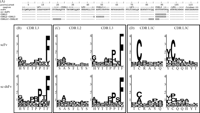
Phage display of antibody fragments from natural or synthetic antibody libraries with the single chain constructs combining the variable fragments (scFv) has been one of the most prominent technologies in antibody engineering. However, the nature of the artificial single chain constructs results in unstable proteins expressed on the phage surface or as soluble proteins secreted in the bacterial culture medium. The stability of the variable domain structures can be enhanced with interdomain disulfide bond, but the single chain disulfide-stabilized constructs (sc-dsFv) have yet to be established as a feasible format for bacterial phage display due to diminishing expression levels on the phage surface in known phage display systems. In this work, biological combinatorial searches were used to establish that the c-region of the signal sequence is critically responsible for effective expression and functional folding of the sc-dsFv on the phage surface. The optimum signal sequences increase the expression of functional sc-dsFv by 2 orders of magnitude compared with wild-type signal sequences, enabling the construction of phage-displayed synthetic antivascular endothelial growth factor sc-dsFv libraries. Comparison of the scFv and sc-dsFv variants selected from the phage-displayed libraries for vascular endothelial growth factor binding revealed the sequence preference differences resulting from the interdomain disulfide bond. These results underlie a new phage display format for antibody fragments with all the benefits from the scFv format but without the downside due to the instability of the dimeric interface in scFv.


Figures

FIGURE 1.
FIGURE 1.
Competitive ELISA for phage-displayed anti-VEGF scFv(fXa+) binding to human VEGF-A. VEGF was immobilized on five sets of wells in a microtiter plate coated with various concentrations of VEGF as follows: 892 nm (black circles), 223 nm (gray circles), 55.8 nm (black upside-down triangles), 13.9 nm (gray triangles), and 3.49 nm (black squares). The x axis shows the concentration of free VEGF added to the corresponding well. The free VEGF molecules compete with the immobilized VEGF for binding to phage-displayed anti-VEGF scFv(fXa+). The phage particles displaying anti-VEGF scFv(fXa+) binding to the immobilized VEGF on the well were quantitatively determined by the ELISA signal shown in the y axis.
FIGURE 2.
FIGURE 2.
Expression level and binding affinity of anti-VEGF sc-dsFv(fXa+) on M13 phage surface or as free soluble protein secreted in the culture medium with the pCANTAB5E phagemid in E. coli strain ER2738. The signal sequence of the pCANTAB5E phagemid is shown in Fig. 3. The x axis shows various single chain antibody variable domain fragment constructs in the pCANTAB5E phagemid. The details of the antibody fragment constructs are described in the text. In the pCANTAB5E phagemid, an E-tag sequence followed by a TAG amber stop codon is encoded between the scFv construct and the pIII sequence (supplemental Fig. 1a). The E-tag was used as a marker for the expression of the scFv/sc-dsFv-pIII fusion protein displayed on the phage surface or as a marker for the free secreted scFv/sc-dsFv molecules. The null control phagemid contains TAA stop codons in the signal sequence (Fig. 3), and thus the phage particles rescued from the E. coli hosts harboring the control phagemid did not display any polypeptide connecting to the pIII minor capsid protein. Also, the bacteria harboring the control phagemid did not secrete free soluble scFv protein. The black histogram and the light gray histogram show the ELISA signals reflecting the binding of the recombinant phage displaying scFv(fXa+)/sc-dsFv(fXa+) to the immobilized anti-E tag antibody (0.1 μg in 100 μl) and to the immobilized VEGF (1 μg in 100 μl), respectively. The overnight culture medium for phage rescue also contained bacteria-secreted soluble protein of the scFv/sc-dsFv constructs, for which the expression stopped at the amber stop codon. The binding of the soluble scFv/sc-dsFv to the anti-E tag antibody and to the VEGF can be detected by HRP-conjugated protein L. Protein L binds to the Vκ light chain of the scFv/sc-dsFv, which in turn binds to immobilized anti-E tag antibody or immobilized VEGF. The dark gray histogram and the white histogram show the ELISA signals reflecting the binding of free soluble scFv/sc-dsFv to the immobilized anti-E tag antibody and to the immobilized VEGF, respectively. The error bars were derived by three repeats of the experiments.
FIGURE 3.
FIGURE 3.
Signal sequence in pCANTAB5E and the constructs of DNA libraries to diversify the tentative signal sequence responsible for the expression of the phage-displayed pIII fusion proteins. The M13pIII-pelB signal sequence for phage-displayed pIII-fusion protein is a combination of the wild-type M13 signal peptide amino-terminal to gene III (MKKLLFAIPLVVPFYSHS) and the pelB signal sequence of P. wasabiae (MKYLLPTAAAGLLLLAAQPAMA). This merged signal sequence (shown in boldface type) was considered containing the tentative n-, h-, and c-regions of the signal sequence. DNA libraries were constructed to diversify the amino acid sequence in the key regions. The primers used in the mutagenesis diversification are shown in the figure. Each of the four of DNA libraries (L1, L2, L4, and L5) contained 10 consecutive NNK (N stands for 25% of G, C, A, and T, and K stands for 50% of G and T; underlined by dashed lines) degenerate codons covering a portion of the tentative signal sequence. Also shown in the figure are the sequences containing TAA stop codons (underlined regions) used as the templates for the library constructions.
FIGURE 4.
FIGURE 4.
Increasing binding to VEGF for signal sequence variants of phage-displayed scFv(fXa+) enriched from the four libraries shown in Fig. 3 after selection/amplification cycles. The complexities of the synthetic phage display library L1, L2, L4, and L5 are 1.4 × 10⁹, 1.3 × 10⁹, 1.2 × 10⁹, and 1.4 × 10⁹, respectively. After each round of selection/amplification cycle, the rescued phage solutions were normalized to 1 × 10¹⁰ cfu/ml, and the binding of the phage particles to immobilized VEGF (each well was coated with 1 μg of VEGF in 100 μl buffer) was measured with ELISA. The ELISA signal strengths are shown in the y axis as functions of the number of the selection/amplification cycles.
FIGURE 5.
FIGURE 5.
Sequence preference patterns emerged from the optimum signal sequences that were selected for optimally facilitating the expression and folding of the phage-displayed anti-VEGF scFv(fXa+). The sequences of the signal peptide emerged from variants selected among a large number of phages enriched for VEGF binding through several cycles (10 cycles for L1 (A) and L4 (C) and 4 cycles for L2 (B) and L5 (D)) of selection/amplification. The top-ranked sequences are listed in supplemental Table 1. The Sequence Logo (42) for these sequences are shown in A–D for variants from the phage display library L1, L2, L4, and L5, respectively.
FIGURE 6.
FIGURE 6.
Effectiveness of the optimum signal sequences from each of the four signal sequence libraries. The optimum signal sequences were selected for enhancing the expression of functional phage-displayed anti-VEGF scFV(fXa+) (Fig. 4). The top-ranked signal sequence variants (-PWLPRDPYIPVVPFYAAQPAMAHHHHHHGH- from L1, -VKKLLPSSLAFLLVFAAQPAMAHHHHHHGH- from L2, -VKKLLFAIPLVVPFYAMSMSRPVASHHHGH- from L4, and -VKKLLFAIPLVVPFYAAQPAYAMSRTPVRS- from L5) were cultured and normalized to 1.0 × 10¹⁰ cfu/ml. The phage solutions were incubated with immobilized VEGF in microtiter wells. The ELISA signals (shown in the y axis) and the titer of the phage particles eluted from the well (shown in the x axis) are plotted for each of the variants. Eight repeats of the experiment were carried out for each of the variants, and the results are shown for the variant from L1 in gray diamonds, L2 in gray circles, L4 in black squares, and L5 in empty triangles. The scales of the ELISA signal and the titer of the bound phage are normalized against the signal and the titer of the phage-displayed anti-VEGF scFv(fXa+) (as shown in empty circles). The scales in both the x axis and the y axis show the folds increased against the data for anti-VEGF scFv(fXa+) encoded in the original pCANTAB5E phagemid with the M13pIII-pelB signal sequence. Both the null control phage and M13KO7 helper phage are negative controls (data shown in black triangles and in black diamonds, respectively). As expected, the data for the negative controls are close to zero in both axes.
FIGURE 7.
FIGURE 7.
Preference sequence pattern for the top-ranked c-region signal sequences enabling the expression of functional phage-displayed anti-VEGF sc-dsFv. The details of the signal sequences, along with the quantitative measurements of the expression efficiency, are listed in supplemental Table 2.
FIGURE 8.
FIGURE 8.
Expression and interface disulfide bond formation in phage-displayed S5 anti-VEGF sc-dsFv(fXa+). The phage-displayed anti-VEGF sc-dsFv was expressed with two L4 signal sequences: SQ(amber stop codon)SMQPSSSL for number 41 and TRSCFAFMLP for number 64 (for more details see supplemental Table 2). All the phage solutions were normalized to 1.0 × 10¹⁰ cfu/ml. One set of the phage solutions was mixed with bovine factor Xa (1 unit) at 37 °C for 1 h (data shown in the gray histogram); the other set of the phage solutions were mixed with only buffer in the same reaction condition (data shown in the black histogram). The binding capabilities of the phage particles from both sets of phage solutions to the immobilized VEGF were measured with ELISA, for which the signal strengths are shown in the y axis. The white histogram shows the binding signal of the phage to immobilized anti-E tag antibody, reflecting the relative expression level of the scFv or sc-dsFv. The error bars were derived from three repeats of the ELISA measurement.
FIGURE 9.
FIGURE 9.
Light chain CDR sequence preferences for VEGF-binding derived from phage-displayed synthetic scFv and sc-dsFv libraries. The light chain sequences of the template scFv and the sc-dsFv are shown in A, along with the randomized CDR regions in the three pairs of synthetic libraries CDRL3, CDRL2-CDRL3, and CDRL1C-CDRL3C. X indicates the locations of the residue encoded with degenerate codon NNK in the synthetic libraries. The complexities for scFv CDRL3 and sc-dsFv CDRL3 library are 9.6 × 10⁸ and 8 × 10⁸, respectively; the complexities for scFv CDRL2-CDRL3 and sc-dsFv CDRL2-CDRL3 library are 1.9 × 10⁹ and 4.6 × 10⁹, respectively; the complexities for scFv CDRL1C-CDRL3C and sc-dsFv CDRL1C-CDRL3C library are 1.3 × 10⁹ and 4.3 × 10⁹, respectively. The signal sequence for both the scFv- and sc-dsFv-pIII fusion proteins is TRSCFAFMLP (see L4 to S5 number 64 shown in Fig. 8, also see supplemental Table 2 for more details). The sequence logos (42) in B–D compare with the sequence preference differences in VEGF binding between the scFv and the sc-dsFv variants from the three pairs of synthetic libraries, respectively. The logos shown in B were derived from 43 and 40 variants for scFv and sc-dsFv, respectively; the logos shown in C were derived from 45 and 42 variants for scFv and sc-dsFv, respectively; the logos shown in D were derived from 38 and 40 variants for scFv and sc-dsFv, respectively. Details of the variants are listed in supplemental Table 3.
FIGURE 10.
FIGURE 10.
Distributions of the VEGF-binding affinity and resistance of fXa digestion for the selected scFv and sc-dsFv variants of which the sequence logos are shown in Fig. 9. In each of the distributions shown in A and B, the boundary of the box closest to zero indicates the 25th percentile; a line within the box marks the median; the boundary of the box farthest from zero indicates the 75th percentile; whiskers (error bars) above and below the box indicate the 90th and 10th percentiles. The dots show the extreme values of the respective distribution. A, the normalized VEGF binding affinity shown in the y axis is defined as follows: normalized VEGF binding = ((VEGFsample − VEGFnull)/(anti_Esampleanti_Enull))/((VEGFWT − VEGFnull)(anti_EWTanti_ Enull), where anti_Esample and VEGFsample are the mean anti-E tag-binding and VEGF-binding ELISA signal, respectively, for the variant in consideration; anti_EWT and VEGFWT (WT is wild type) are the mean anti-E tag-binding and VEGF-binding ELISA signal, respectively, for the template anti-VEGF scFv/sc-dsFv, for which the sequences for the CDR regions are shown in Fig. 9A. The corresponding null values (anti_Enull and VEGFnull) were derived with a negative control phage without the displayed fusion protein. B, VEGF binding % after fXa treatment shown in the y axis is defined as follows: VEGF binding % = ((VEGFsamplefXa − VEGFnullfXa)/(VEGFsample − VEGFnull)) × 100%, where VEGFsample and VEGFsamplefXa are the mean VEGF-binding ELISA signal before and after fXa treatment for the variant in consideration. The corresponding null values (VEGFnull and VEGFnullfXa) were derived with a negative control phage without the displayed fusion protein.

Similar articles

Cited by

References

    1. Yokota T., Milenic D. E., Whitlow M., Schlom J. (1992) Cancer Res. 52, 3402–3408 - PubMed
    1. Michnick S. W., Sidhu S. S. (2008) Nat. Chem. Biol. 4, 326–329 - PubMed
    1. Wark K. L., Hudson P. J. (2006) Adv. Drug Deliv. Rev. 58, 657–670 - PubMed
    1. Carter P. J. (2006) Nat. Rev. Immunol. 6, 343–357 - PubMed
    1. Hoogenboom H. R. (2005) Nat. Biotechnol. 23, 1105–1116 - PubMed

Publication types

MeSH terms

LinkOut - more resources