. 2010 Mar 12;285(11):7964-76.
doi: 10.1074/jbc.M109.063586. Epub 2010 Jan 12.

Alkaline ceramidase 3 (ACER3) hydrolyzes unsaturated long-chain ceramides, and its down-regulation inhibits both cell proliferation and apoptosis

Affiliations

Alkaline ceramidase 3 (ACER3) hydrolyzes unsaturated long-chain ceramides, and its down-regulation inhibits both cell proliferation and apoptosis

Wei Hu et al. J Biol Chem. .

Abstract

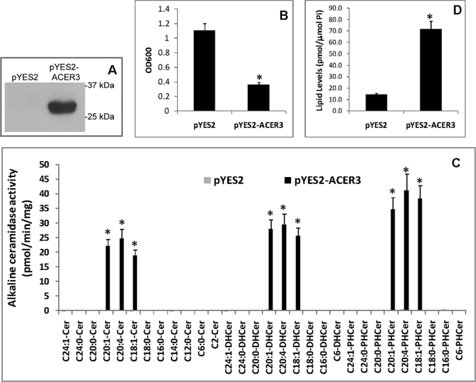
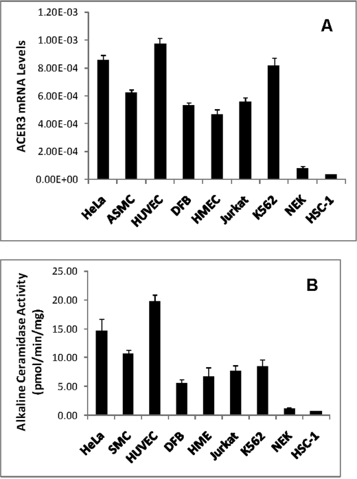
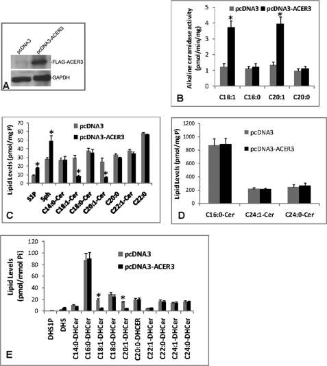
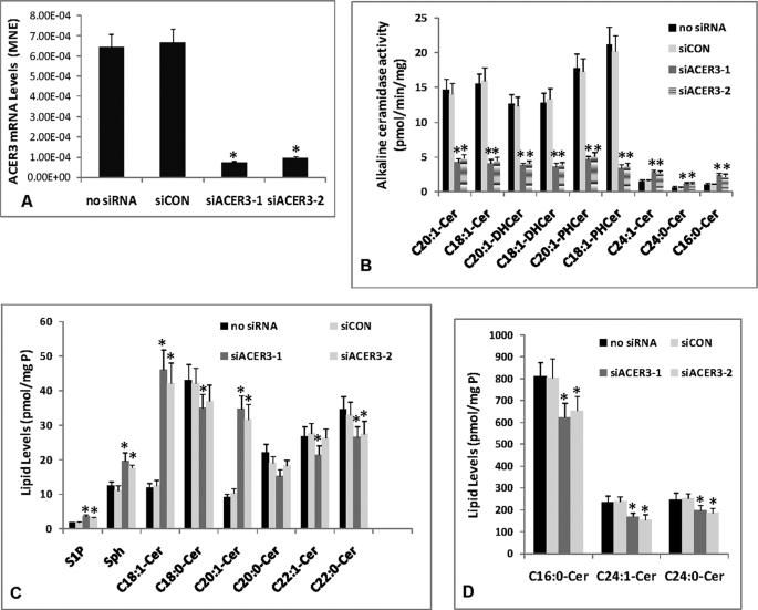
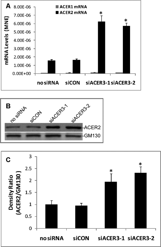
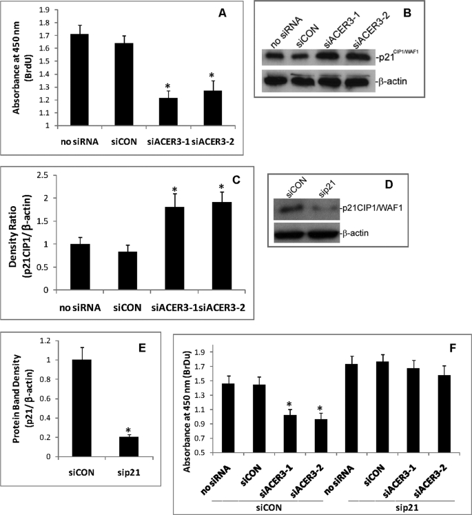
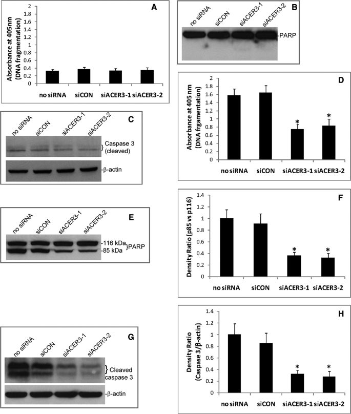
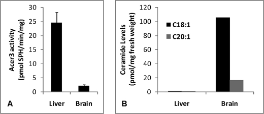
Ceramides with different fatty acyl chains may vary in their physiological or pathological roles; however, it remains unclear how cellular levels of individual ceramide species are regulated. Here, we demonstrate that our previously cloned human alkaline ceramidase 3 (ACER3) specifically controls the hydrolysis of ceramides carrying unsaturated long acyl chains, unsaturated long-chain (ULC) ceramides. In vitro, ACER3 only hydrolyzed C(18:1)-, C(20:1)-, C(20:4)-ceramides, dihydroceramides, and phytoceramides. In cells, ACER3 overexpression decreased C(18:1)- and C(20:1)-ceramides and dihydroceramides, whereas ACER3 knockdown by RNA interference had the opposite effect, suggesting that ACER3 controls the catabolism of ULC ceramides and dihydroceramides. ACER3 knockdown inhibited cell proliferation and up-regulated the cyclin-dependent kinase inhibitor p21(CIP1/WAF1). Blocking p21(CIP1/WAF1) up-regulation attenuated the inhibitory effect of ACER3 knockdown on cell proliferation, suggesting that ACER3 knockdown inhibits cell proliferation because of p21(CIP1/WAF1) up-regulation. ACER3 knockdown inhibited cell apoptosis in response to serum deprivation. ACER3 knockdown up-regulated the expression of the alkaline ceramidase 2 (ACER2), and the ACER2 up-regulation decreased non-ULC ceramide species while increasing both sphingosine and its phosphate. Collectively, these data suggest that ACER3 catalyzes the hydrolysis of ULC ceramides and dihydroceramides and that ACER3 coordinates with ACER2 to regulate cell proliferation and survival.


Figures

FIGURE 1.
FIGURE 1.
ACER3 hydrolyzes C18:1-, C20:1-, and C20:4-(phyto/dihydro)ceramides specifically. A, Δypc1Δydc1YES2 (pYES2) or Δypc1Δydc1ACER3 (pYES2-ACER3) cells were grown in galactose-containing medium (SD-gal/raf) before microsomes were isolated from these cells. Proteins were solubilized with 1% Triton X-100 from the microsomes and were analyzed by Western blot using anti-FLAG antibody at a 1:1000 dilution. Each lane contained 40 μg of proteins. B, Δypc1Δydc1YES2 or Δypc1Δydc1ACER3 cells were grown to the stationary phase in glucose-containing medium (SD-glu) before being diluted to SD-gal/raf medium. Yeast cells were then grown in SD-gal/raf medium for 16 h before cell density was determined by absorbance at 600 nm (A600). Note that the growth of Δypc1Δydc1ACER3 cells was significantly inhibited compared with the growth of Δypc1Δydc1YES2 cells. C, Δypc1Δydc1YES2 or Δypc1Δydc1ACER3 cells grown in SD-glu medium were switched to SD-gal/raf medium. At 8 h post-medium switch, yeast cells were harvested, and microsomes were prepared. Microsomal alkaline ceramidase activity was determined at pH 9.0 using various ceramides (Cer), dihydroceramides (DHCer), and phytoceramides (PHCer) as substrates. Each enzymatic reaction contained 150 μm substrate and ∼40 μg of proteins. D, lipids were extracted from Δypc1Δydc1YES2 or Δypc1Δydc1ACER3 cells grown in SD-gal/raf medium according to the Bligh-Dyer method (27), and PHS levels were determined by HPLC. PHS levels were normalized to total inorganic phosphate (Pi) in lipid extracts. Numerical data represent mean values ± S.D. of three independent experiments. Image datum represents one of three independent experiments with similar results. *, p < 0.05 versus control, n = 3.
FIGURE 2.
FIGURE 2.
ACER3 mRNAs are highly expressed in various cell types. A, total RNA was isolated from different cell types, and ACER3 mRNA levels were determined by qPCR analysis. B, microsomes were isolated from indicated cell types, and alkaline ceramidase activity in the microsomes was measured using d-ribo-C12-NBD-phytoceramide (100 μm) as substrate. ASMC, aortic smooth muscle cells; DFC, dermal fibroblast cells; HMEC, human mammary epithelial cells; and NEK, normal human epidermal keratinocytes. Data represent mean values ± S.D. of three independent experiments.
FIGURE 3.
FIGURE 3.
ACER3 overexpression decreases ULC ceramides while increasing SPH and S1P in cells. A, HSC-1 cells were transiently transfected with pcDNA3 or pcDNA3-ACER3 using Effectene according to the manufacturer's instructions. At 72 h post-transfection, microsomes were prepared from cells. Proteins (80 μg) solubilized from a portion of each microsomal preparation were analyzed by Western blot using an anti-FLAG antibody at a 1:1000 dilution. GAPDH, glyceraldehyde-3-phosphate dehydrogenase. B, remaining portion of each microsomal preparation from A was measured for alkaline ceramidase activity at pH 9.0 and with the indicated ceramides (150 μm) as substrates. C–E, HSC-1 cells transected with pcDNA3 or pcDNA3-ACER3 were analyzed by ESI/MS/MS for sphingolipids. Sphingolipid levels were normalized to total phospholipids (Pi). Numerical data represent mean values ± S.D. of three independent experiments. Image datum represents one of two independent experiments with similar results. *, p < 0.05 versus control.
FIGURE 4.
FIGURE 4.
ACER3 knockdown by RNAi increases ULC ceramides in cells. A–D, HeLa cells at a 50% confluence were transfected with 5 nm siCON, siACER3-1, or siACER3-2, or with buffer only (no siRNA). At 48 h post-siRNA transfection, cells were harvested for qPCR analysis of ACER3 mRNA levels (A), alkaline ceramidase activity assays (B), or ESI/MS/MS analyses of sphingolipids (C and D). Data represent mean values ± S.D. of three independent experiments. *, p < 0.05 versus the control siCON or no siRNA. MNE, mean normalized expression.
FIGURE 5.
FIGURE 5.
ACER3 knockdown up-regulates ACER2. A–C, HeLa cells were transfected with siCON, siACER3-1, or siACER3-2 or siACER3 at 5 nm or with no siRNA for 48 h before qPCR analysis for ACER1 or ACER2 mRNA levels (A), Western blot analysis for ACER2 and β-actin (B), or densitometry for ACER2 and β-actin (C) was performed. Densitometry was performed on a ChemiImager 4400 system (Alpha Innotech, San Leandro, CA). The ACER2/β-actin density ratio in cells transfected with no siRNA was arbitrarily set as 1, and the relative ACER2/β-actin density ratios were accordingly computed in cells transfected with each of other different siRNAs. Data represent mean values ± S.D. of three independent experiments. Image datum represents one of three independent experiments with similar results. *, p < 0.05 versus the controls (siCON or no siRNA). MNE, mean normalized expression.
FIGURE 6.
FIGURE 6.
ACER3 knockdown inhibits cell proliferation. A, HeLa cells were transfected with siCON, siACER3-1, siACER3-2, or with no siRNA as described in Fig. 5 for 72 h before cell proliferation ELISA was performed. B and C, HeLa cells were transfected with each of the indicated siRNAs for 48 h before p21CIP1/WAF1 levels were analyzed by Western blot (B) and quantified by densitometry (C). D and E, HeLa cells were transfected with siCON (5 nm) or sip21 (5 nm), a sip21CIP1/WAF1-specific siRNA for 48 h before the content of p21CIP1/WAF1 was analyzed by Western blot with anti-p21CIP1/WAF1 antibody (D), followed by densitometry (E). F, HeLa cells were transfected with siCON plus siACER3-1, siCON plus siACER3-2, siCON plus buffer only (no siRNA), sip21 plus siCON, sip21 plus siACER3-1, sip21 plus siACER3, or sip21 plus buffer only (no siRNA) at 5 nm each. At 72 h post-siRNA transfection, cells were subjected to cell proliferation ELISA. Numerical data represent mean values ± S.D. of three independent experiments. Image datum represents one of three independent experiments with similar results. *, p < 0.05 versus the control (siCON or no siRNA).
FIGURE 7.
FIGURE 7.
ACER3 knockdown inhibits apoptosis in response to serum deprivation. A–C, HeLa cells were transfected with indicated siRNAs as described in Fig. 5 in regular medium containing 10% fetal bovine serum for 72 h before they were subjected to cell death detection ELISA (A), Western blot analysis for PARP cleavage (B), or caspase 3 cleavage (C). D–F, HeLa cells were transfected with indicated siRNAs for 24 h before they were switched to serum-free medium. At 48 h post-serum deprivation, the cells were subjected to cell death detection ELISA (D), Western blot analysis for PARP cleavage (E), densitometry of PARP (F), Western blot analysis for caspase 3 cleavage (G), and densitometry of caspase 3 (H). Numerical data represent mean values ± S.D. of three independent experiments. Image data represent one of three independent experiments with similar results. *, p < 0.05 versus the control (siCON or no siRNA).
FIGURE 8.
FIGURE 8.
ULC ceramides are abundant in the mouse brain with low ACER3 activity. A 2-month old male mouse was sacrificed, and liver and brain tissues were resected. The same fresh weight of liver and brain tissues were homogenized in 25 mm Tris-HCl (pH 7.4) containing EDTA (5 mm), EGTA (5 mm), and NaCl (150 mm). Tissue homogenates of each tissue were divided into 4 aliquots with the same volume. Two aliquots were assayed for ACER3 activity on NBD-C12-phytoceramide (A), and the remaining aliquots were analyzed by ESI/MS/MS for the of C18:0- and C20:1-ceramides (B).

References

    1. Mao C., Obeid L. M. (2008) Biochim. Biophys. Acta 1781, 424–434 - PMC - PubMed
    1. Hanna A. N., Berthiaume L. G., Kikuchi Y., Begg D., Bourgoin S., Brindley D. N. (2001) Mol. Biol. Cell 12, 3618–3630 - PMC - PubMed
    1. Charles A. G., Han T. Y., Liu Y. Y., Hansen N., Giuliano A. E., Cabot M. C. (2001) Cancer Chemother. Pharmacol. 47, 444–450 - PubMed
    1. Magnoni C., Euclidi E., Benassi L., Bertazzoni G., Cossarizza A., Seidenari S., Giannetti A. (2002) Toxicol. In Vitro 16, 349–355 - PubMed
    1. Jaffrézou J. P., Bruno A. P., Moisand A., Levade T., Laurent G. (2001) FASEB J. 15, 123–133 - PubMed

Publication types

MeSH terms