. 2010 Jan;8(1):93-106.
doi: 10.1158/1541-7786.MCR-08-0491. Epub 2010 Jan 12.

The TRC8 ubiquitin ligase is sterol regulated and interacts with lipid and protein biosynthetic pathways

Affiliations

The TRC8 ubiquitin ligase is sterol regulated and interacts with lipid and protein biosynthetic pathways

Jason P Lee et al. Mol Cancer Res. 2010 Jan.

Abstract

TRC8/RNF139 encodes an endoplasmic reticulum-resident E3 ubiquitin ligase that inhibits growth in a RING- and ubiquitylation-dependent manner. TRC8 also contains a predicted sterol-sensing domain. Here, we report that TRC8 protein levels are sterol responsive and that it binds and stimulates ubiquitylation of the endoplasmic reticulum anchor protein INSIG. Induction of TRC8 destabilized the precursor forms of the transcription factors SREBP-1 and SREBP-2. Loss of SREBP precursors was proteasome dependent, required a functional RING domain, occurred without generating processed nuclear forms, and suppressed SREBP target genes. TRC8 knockdown had opposite effects in sterol-deprived cells. In Drosophila, growth inhibition by DTrc8 was genetically suppressed by loss of specific Mprlp, Padlp N-terminal domain-containing proteins found in the COP9 signalosome and eIF3. DTrc8 genetically and physically interacted with two eIF3 subunits: eIF3f and eIF3h. Coimmunoprecipitation experiments confirmed these interactions in mammalian cells, and TRC8 overexpression suppressed polysome profiles. Moreover, high-molecular weight ubiquitylated proteins were observed in eIF3 immunoprecipitations from TRC8-overexpressing cells. Thus, TRC8 function may provide a regulatory link between the lipid and protein biosynthetic pathways.


Figures

Figure 1
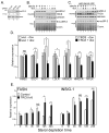
Figure 1. TRC8 levels are modulated by sterols
(A) CHO FlpIn TRex cells containing dox-inducible TRC8-HA were cultured in the absence of dox with either abundant sterols (+ sterols; 5% FCS in F-12) or without (− sterols; 5% LPDS, 50 μM mevastatin, 50 μM mevalonate). The indicated time points were harvested, lysed and 10 μg aliquots analyzed by Western blot for TRC8-HA. Controls included HMGCR and actin. (B) HEK293 cells stably transfected with the TRC8-HA-FlpIn construct were cultured in sterol-depletion media for 18h to accumulate TRC8. Excess sterols were then added (+ sterols; 10μg/mL 25-HC and 50 μg/mL cholesterol) to all but the control (0h) in the absence of cycloheximide (CHX), and time points analyzed for decay of TRC8-HA and HMGCR; GAPDH indicates loading. (C) HEK293 cells containing wild type TRC8-HA or the RING mutant, C547S;C550S, were dox-induced for 24 h then sterol-starved for 1h in DMEM with 5% LPDS and 1% hydroxypropyl β-cyclodextrin (HPβCD). Media were then changed to DMEM with 5% LPDS containing either 10 μM statin and 50 μM mevalonate (sterols) or 50 μg/mL cholesterol/10 μg/mL 25-HC (+ sterols). CHX was then added to 10 μM and time points harvested at 3h intervals for analysis by Western blot using HA and actin antibodies. (D) Fluorescent band intensities from triplicate samples as in (C) were analyzed on an Odyssey IR fluorescence scanner and quantified. Points represent means, +/− s.d., from three replicates; gels and graphs are representative of 3 independent experiments.
Figure 2
Figure 2. Sterols regulate endogenous TRC8 and induce ubiquitylation
(A) Membranes purified from HEK293 cells stably transfected with a tet-inducible shRNA targeting TRC8 permitted detection of endogenous TRC8 by Western blot (lanes 1, 2, −dox). Identity of the band was confirmed by 4 days of dox treatment (+dox) to induce knockdown. Parallel cultures were sterol-starved for 24 (lanes 5–8) or 48h (lanes 9–12) and membrane preparations analyzed for endogenous TRC8 and HMGCR (postive control). Coomassie staining verified equal loading. (B) Data for 24 h sterol starvation were densitized and quantified. Each bar represents the mean of TRC8 signal in triplicate samples, +/− s.d. RNA isolated from the same cultures was analyzed by realtime RT-PCR for endogenous TRC8 and INSIG-1 mRNAs. Delta Ct values were converted to fold expression and normalized to control samples (+sterols). (C) TRC8-HA or control (vec) HEK293 cells, grown in sterol-replete medium, were transfected with flag-tagged ubiquitin (1 μg), dox-induced and harvested 24 h later following MG132 addition (10 μM) during the final 2h. Lysates (200 μg aliquots) were immunoprecipitated with anti-HA or murine IgG (controls) and IP pellets analyzed on Western blots for flag-ubiquitin. HA immunoblots showed equal recovery of TRC8-HA; this and other HA blots were cropped to remove strong IgG background. Actin and HA blots verified equal input. (D) TRC8-HA and control cells were transfected with flag-ubiquitin and induced with dox as in (C). Cells were then sterol-starved for 15h followed by addition of cholesterol (12.5 μM) and 25-HC (10 μg/mL) for 3h to the indicated plates. MG132 was added 2h prior to harvest and lysates (200 μg aliquots) were immunoprecipated with anti- HA beads. Western blot analysis utilized the indicated antibodies.
Figure 3
Figure 3. TRC8 binds and ubiquitylates INSIG
(A) TRC8-HA-FlpIn cells (wt or C547S;C550S RING mutant), cultured in sterol-replete DMEM (10% FCS), were transfected overnight with 300ng of pCMV-INSIG-1-myc or pCMV-INSIG-2-myc, then dox-treated or not for 24 h. Where indicated, MG132 was added 4h prior to harvest. CHAPSO lysates (500μg) were immunoprecipitated with anti-HA and pellets analyzed by Western blot for INSIG (myc) and TRC8-HA (HA). (B) Diagram of TRC8 showing 10 TM segments (bars), the first five of which comprise the SSD (black bars). TRC8 also contains a RING-H2 domain in the C-terminus (shaded circle). The conserved tyrosine 32, in the first TM of the SSD, was mutated to glutamate or cysteine and used for the IP experiments in panel C. (C) Control HEK293 cells (vec) and stable transfectants expressing wild type or SSD mutant TRC8 (Y32E or Y32C) were transfected with INSIG-1-myc and harvested after 48h. TritonX-100 lysates containing 400ug of protein were immunoprecipitated using anti-HA or control beads. IP pellets were analyzed for co-precipitated INSIG-1 (myc) and TRC8 (HA). Input blots verified equal expression; asterisks indicate background IgG bands. (D) Control cells (vec) and stable transfectants expressing wild type or RING mutant TRC8 (C547S, C550S) were co-transfected with flag-ubiquitin and myc-INSIG-1 (wt) or the K156R, K158R mutant (kk), as indicated. Cells were dox-induced for 24 h and treated with MG132 for 2h prior to harvest. TritonX-100 lysates (200 μg aliquots) were immunoprecipitated with anti-flag beads and analyzed on Western blots with the indicated antibodies. Input blots verified equal expression; conjugated ubiquitin, detected with anti-flag antibodies, was used to verify equal expression because unconjugated ubiquitin was not visible.
Figure 4
Figure 4. TRC8 destablizes SREBP precursors in a RING and proteasome-dependent manner
(A) TRC8-HA and control HEK293 cells (vector) were treated for 24 h with increasing dox (ng/ml). RIPA buffer lysates (10 μg/lane) were analyzed for preSREBP-1 (2A4 antibody), preSREBP-2 (1C6 antibody) and TRC8-HA; Coomassie stain verified equal loading. (B) HEK293 cells containing TRC8-HA or three RING mutations (ΔRING, C547S;C550S and S557A;R559A) were induced for 24 h with increasing doses of dox (ng/ml) and analyzed as in (A). Ubiquitylation activity is only present in wild type TRC8 and in the S557A;R559A mutation, as indicated (19). The ΔRING mutation removes 39 amino acids, or about 4kDa, and detectably increases mobility on SDS/PAGE. (C) Duplicate plates of TRC8-HA or control HEK293 cells were dox-induced (100 ng/ml) for 12 h, then MG132 (10 μM) was added as indicated. After 9 h, cells were harvested and analyzed for the indicated proteins. HIF1α accumulation verified proteasome inhibition by MG132 (2); actin verified loading. (D) Duplicate plates of TRC8-HA or vector HEK293 cells were cultured in excess sterols (12.5 μM cholesterol, 10 μg/mL 25-HC), with or without dox, as indicated. After 24 h, cells were processed into membrane and nuclear fractions [Sever et al., (2003) (29), see Methods]. Fractions were analyzed for precursor and nuclear forms of SREBP-1/2 using 2A4 (SREBP-1) and 1D2 (SREBP-2) antibodies.
Figure 5
Figure 5. TRC8 knockdown upregulates SREBP-2
(A) HEK293 cells cultured in DMEM (10% FCS) were transfected with the indicated siRNAs (mock, scrambled or TRC8-specific [Ambion #-136327]). Detergent lysates were analyzed 72h post-transfection for pre-SREBP-2 and actin. Densitometry was performed on serial dilutions of replicate (n = 6) transfected lysates, relative expression values are means, +/− s.d. Representative blots are shown. (B) Duplicate cultures of HEK293 cells stably transfected with the inducible shRNA-TRC8 construct were treated for 4 days with vehicle (−) or dox (+), as indicated, to knockdown endogenous TRC8 (Fig. S2C). Cells were then incubated with fresh 5% FCS (+ sterols) or sterol-depletion media (sterols) for 20 h, harvested and processed into membrane and nuclear fractions. Western blots were analyzed for precursor and nucSREBP-2 (1D2); calnexin and Coomassie stain provided loading controls; knockdown of TRC8 was verified using anti-RNF139 antibody. (C) Cell cultures identical to (B) were treated with excess sterols (12.5 μg/mL cholesterol/10 μg/mL 25-HC) for the final 6h prior to harvest to arrest processing and accumulate SREBP precursors. Membrane and nuclear fractions were analyzed for SREBP-2 (1D2); knockdown of TRC8 was verified using anti-RNF139 antibody. Calnexin and a Coomassie stained gel provided loading controls. (D) Cultures of TRC8-HA or control (vec) HEK293 cells in DMEM (10% FCS) were treated with vehicle or dox (100 ng/mL) to induce TRC8-HA and harvested for RNA 24 h later. qRT-PCR was conducted for the indicated genes. Ct values were normalized geometric means of 4 controls; HMBS is graphed as a negative control. Bars represent means +/− SEM; *p < 0.05; **p < 0.01 by Student’s t-test (n = 6). Vector – dox (open bars); vector + dox (grey bars); TRC8-HA – dox (striped bars); TRC8-HA + dox (black bars). (E). TRC8 knockdown cells, treated with dox as in (B, C) to reduce TRC8, were sterol-deprived for the indicated times. Quadruplicate samples were analyzed by qRT-PCR for SREBP target genes. Bars represent means +/− SEM; *p < 0.05; **p < 0.01 by Student’s t-test (n = 4). TRC8 k/d cells – dox (open bars); + dox (black bars).
Figure 6
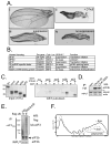
Figure 6. Genetic and physical interaction of TRC8 with subunits of eIF3
(A; i–iv) Examples of male fruit fly wings; (i) wild type; (ii) DTrc8 over-expression; (iii) suppressor locus with heterozygous loss-of-function mutation (partially restores growth to DTrc8-inhibited wing); (iv) equivalent mutation in non-suppressor locus (fails to restore growth). (B) Results of suppressor screen among 6 genes containing MPN domains. All three loci encoding subunits of eIF3 suppressed the DTrc8 wing phenotype. Note there are two eIF3f homologs in flies. (C) GST pull-down assays of labeled TNT products. Purified GST and GST-DTrc8562–809 fusion proteins (21) were mixed with [35S] met-labeled in vitro translation products, as indicated. Bound proteins were identified by SDS-PAGE and autoradiography. Negative controls included luciferase (luc) and the proteasome subunit Rpn8 (21); CSN5 provided a positive control. The eIF3f isoform shown derived from the Drosophila gene CG9769. (D) HEK293 cells carrying TRC8-HA, vector or RING-mutant (C547S;C550S) were dox-induced for 24 h. Lysates were immunoprecipitated with anti-HA beads and bound proteins analyzed by Western blot for eIF3b, a core eIF3 subunit. Tubulin and actin established equal inputs. (E) TRC8-HA and control 293 cells were transfected with flag-ubiquitin (1 μg) and dox-induced for 24 h. Cells were harvested following MG132 addition (10 μM) during the final 2h. Triton X 100 lysates (200 μg aliquots) were immunoprecipitated with antibodies directed against the eIF3 holocomplex (54). IP pellets were analyzed by Western blot for flag-ubiquitin conjugated proteins and eIF3b as an indicator of eIF3 complex recovery and for equal input. (F) Polysome suppression by TRC8. TRC8-HA cells were treated with dox or vehicle for 24 h and harvested (see Methods). Polysomes were analyzed on 10 to 50% sucrose density gradients (53) to generate A280 gradient profiles; the first three polysome peaks are labeled 1–3. Results shown are representative of three repetitions.
Figure 7
Figure 7. Model for TRC8 interaction with sterols and protein translation
(A) Cells cultured in media with 10% FCS (normal lipoprotein/sterol content) undergo basal preSREBP processing (dashed arrows) and contain low levels of TRC8, which has only modest effects on growth, translation and SREBPs (indicated by the dashed lines). (B) Acute removal of sterols leads to rapid activation of preSREBPs with increases in SREBP target gene expression, including SREBPs themselves in a positive feed-forward loop (bold arrows). Production of lipid biosynthetic enzymes initiates restoration of lipid homeostasis. Initially, TRC8 does not restrain this process due to low levels, but the accumulation process begins so that by 12 to 24h of sterol deprivation (panel C), TRC8 causes a reduction of growth, reduction of translation and specific reduction of SREBPs (and other ER proteins like INSIG). This action may limit the stress of over-accumulating transmembrane proteins in an ER compartment compromised by loss of sterols and other lipids. Normalization of lipid levels occurs because TRC8-mediated reductions are not complete, permitting lipid synthesis to continue even as membrane protein accumulation becomes restricted. Moreover, reduced proliferative rates caused by accumulating TRC8 would permit restoration of lipid homeostasis by reducing requirements for membrane biogenesis. Restored lipids would then destabilize TRC8, releasing its inhibition of growth and translation.

Similar articles

Cited by

References

    1. Jemal A, Siegel R, Ward E, et al. Cancer statistics. CA Cancer J Clin. 2006;56(2):106–30. - PubMed
    1. Kaelin WG., Jr The von Hippel-Lindau protein, HIF hydroxylation, and oxygen sensing. Biochemical and biophysical research communications. 2005;338(1):627–38. - PubMed
    1. Hughes MD, Kapllani E, Alexander AE, Burk RD, Schoenfeld AR. HIF-2alpha downregulation in the absence of functional VHL is not sufficient for renal cell differentiation. Cancer cell international. 2007;7:13. - PMC - PubMed
    1. Mandriota SJ, Turner KJ, Davies DR, et al. HIF activation identifies early lesions in VHL kidneys: evidence for site-specific tumor suppressor function in the nephron. Cancer cell. 2002;1(5):459–68. - PubMed
    1. Tomlinson IP, Alam NA, Rowan AJ, et al. Germline mutations in FH predispose to dominantly inherited uterine fibroids, skin leiomyomata and papillary renal cell cancer. Nature genetics. 2002;30(4):406–10. - PubMed

Publication types

MeSH terms