. 2010 Jan 15;16(2):600-9.
doi: 10.1158/1078-0432.CCR-09-1979. Epub 2010 Jan 12.

Secretory leukocyte protease inhibitor antagonizes paclitaxel in ovarian cancer cells

Affiliations

Secretory leukocyte protease inhibitor antagonizes paclitaxel in ovarian cancer cells

Nabila Rasool et al. Clin Cancer Res. .

Abstract

Purpose: Ovarian cancer recurrence with the development of paclitaxel resistance is an obstacle to long-term survival. We showed that secretory leukocyte protease inhibitor (SLPI) is a survival factor for ovarian cancer. We hypothesize that SLPI may antagonize paclitaxel injury.

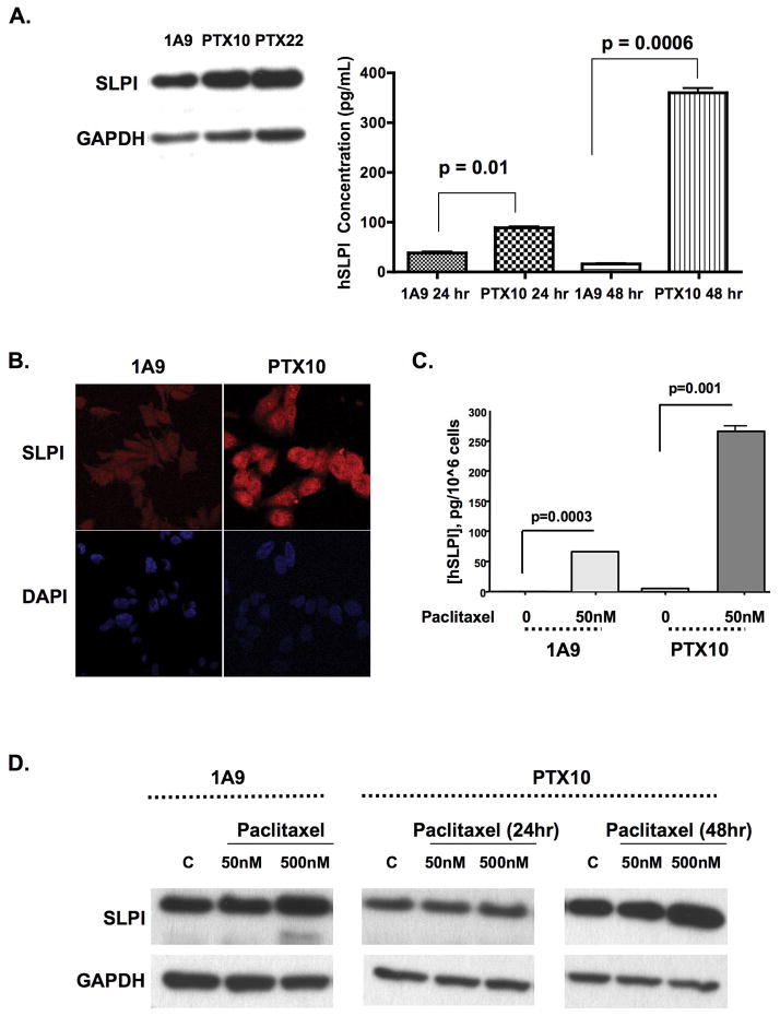
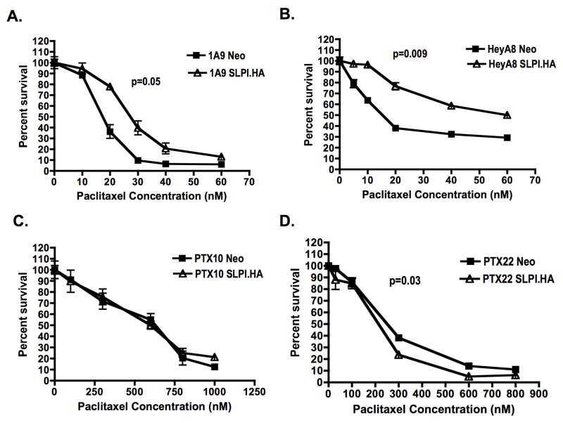
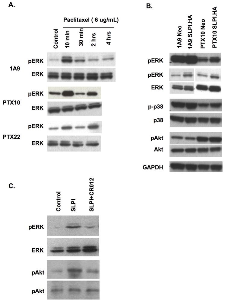
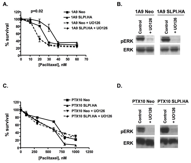
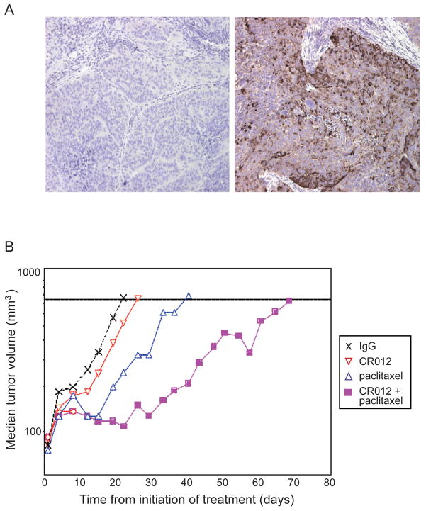
Experimental design: Differential SLPI induction in response to paclitaxel and in response to stable forced expression of SLPI was shown in A2780-1A9 cells and their paclitaxel-resistant sublines, PTX10 and PTX22, and confirmed with HEY-A8 cells. SLPI-mediated survival was reduced by the MAP/extracellular signal-regulated kinase (ERK) kinase inhibitor, U0126, and a humanized neutralizing monoclonal anti-SLPI antibody, CR012. OVCAR3 xenographs tested the role of CR012 in vivo.

Results: SLPI expression was lower in A2780-1A9 ovarian cancer cells than in PTX10 and PTX22, and SLPI was induced by paclitaxel exposure. Stable SLPI expression yielded a proliferation advantage (P = 0.01); expression of and response to SLPI in OVCAR3 cells were abrogated by exposure to CR012. SLPI reduced the paclitaxel susceptibility of 1A9 and HEY-A8 cells (P <or= 0.05), and SLPI expression did not increase the resistance of PTX10 and PTX22 cells. Both paclitaxel and SLPI overexpression induced ERK activation. Inhibition of MAP/ERK kinase with U0126 increased paclitaxel injury and overcame SLPI-mediated cell protection. It did not reinstate PTX10 sensitivity to paclitaxel, which was associated with AKT activation. Significant inhibition of OVCAR3 xenograft growth was observed with CR012 and paclitaxel, over single agents (P <or= 0.001).

Conclusions: A two-pronged approach confirmed that SLPI overcomes paclitaxel in part through activation of ERK1/2. These results credential SLPI as a molecular target for ovarian cancer and suggest CR012 as a tool for proof of concept.


Figures

Figure 1
Figure 1. SLPI increases cell proliferation
A. Forced expression of HA-tagged SLPI stimulates growth. Inset shows HA tag expression. P-values represent the statistical significance for the entire curve. B. CR012 recognizes SLPI. CR012 is credentialed by its recognition SLPI by immunoprecipitation/immunoblot in OVCAR3 lysates (lanes: 1-SLPI antisense-treated cells, 2-scramble-treated, 3-transfection medium control, 4-no exposure), and it illuminates membrane-bound SLPI (B; blue: untreated; green: IgG2 control; red: CR012, 2ug/mL). C, D. CR012 is a neutralizing anti-SLPI antibody. CR012 blocks SLPI inhibition of elastase activity in vitro (C) and reduces proliferation in the presence or absence of added SLPI (D). IgG2 isotype control is used to show specificity. Representative data of at least three independent experiments are shown.
Figure 2
Figure 2. Paclitaxel exposure and resistance stimulate SLPI in ovarian cancer cells
SLPI is upregulated in paclitaxel-resistant cells. (A) Increased SLPI production and secretion in lysates (immunoblot) and CM (ELISA) in samples collected after 24hr serum starvation. Immunofluorescence (B) confirms upregulation and similar cellular localization. C, D. Exposure to paclitaxel stimulates cellular SLPI (D, immunoblot) and CM (E, ELISA). PTX10 cells were exposed to 50 or 500nM paclitaxel over 24 and 48hr; 1A9 cells were only treated for 24hr. Conditioned medium SLPI concentration is expressed as pg/106 cells to normalize for cell number. Representative data of at least three independent experiments are shown.
Figure 3
Figure 3. Overexpression of SLPI confers paclitaxel resistance to wild type but not paclitaxel-resistant cells
Cells were pulsed with paclitaxel at 0–70nM (A, B) or 1000nM (C, D) for 6hr then allowed to grow for 4d. (A) 1A9; (B) HeyA8; (C) PTX10; (D) PTX22. Representative data of at least three independent experiments are shown. P-values represent the statistical significance for the entire curve.
Figure 4
Figure 4. Both paclitaxel treatment and transfection of SLPI.HA activate ERK
A. Paclitaxel pulse induces phospho-ERK. Paclitaxel (6ug/mL) was pulsed over 10min to 4hr prior to cell harvesting. A biphasic activation is observed in the resistant cells. B. Paclitaxel and SLPI induce ERK activation, but only paclitaxel resistance is associated with activation of AKT in the A2780 cell set. Two panels of pERK are presented, independent experiments, one showing a darker view and one a shorter exposure. Representative data of at least three independent experiments are shown. GAPDH represents the loading control. C. CR012 reduces activation of ERK and AKT.
Figure 5
Figure 5. The MAPK inhibitor, UO126, reverses the SLPI-mediated paclitaxel resistance only in wild type cells
A, C. Paclitaxel survival curves and downregulation of pERK with U0126 (10μM) in 1A9 or PTX10 cells. Neo and SLPI.HA cells were pretreated with UO126 for 1hr then UO126 was maintained throughout the assay. After the first hour, cells had a 6hr pulse of paclitaxel; the XTT assay was performed 4d later. B, D. Immunoblot demonstration of loss of pERK activation. Western immunoblot demonstrates ERK and pERK expression in cells treated with UO126 for 24hr in serum-free medium. Representative data of at least three independent experiments are shown. P-values represent the statistical significance for the entire curve.
Figure 6
Figure 6. CR012 augments antiproliferative effects of paclitaxel in xenografts
A. CR012 recognizes SLPI expression in OVCAR3 xenografts. Immunohistochemical staining with CR012 (right) or IgG2 isotype control (left) indicates specific staining. B. CR012 augments anticancer activity of paclitaxel. Median tumor volumes are plotted as a function of time. The combination of CR012 and paclitaxel yielded a T/C of 298% (p<0.001). Treatment started on day 1: IgG2 or CR012, 1 mg/kg by tail vein every 4 days × 4 doses; paclitaxel 7.5 mg/kg by tail vein every other day × 5 doses.

Similar articles

Cited by

References

    1. Jemal A, Siegel R, Ward E, et al. Cancer statistics. CA: a cancer journal for clinicians. 2008;58:71–96. - PubMed
    1. Einzig AI, Wiernik PH, Sasloff J, Runowicz CD, Goldberg GL. Phase II study and long-term follow-up of patients treated with taxol for advanced ovarian adenocarcinoma. J Clin Oncol. 1992;10:1748–53. - PubMed
    1. du Bois A, Luck HJ, Meier W, et al. A randomized clinical trial of cisplatin/paclitaxel versus carboplatin/paclitaxel as first-line treatment of ovarian cancer. J Natl Cancer Inst. 2003;95:1320–9. - PubMed
    1. Agarwal R, Kaye SB. Ovarian cancer: strategies for overcoming resistance to chemotherapy. Nat Rev Cancer. 2003;3:502–16. - PubMed
    1. Hough CD, Cho KR, Zonderman AB, Schwartz DR, Morin PJ. Coordinately up-regulated genes in ovarian cancer. Cancer Res. 2001;61:3869–76. - PubMed

Publication types

MeSH terms

Substances