. 2010 Jan 15;16(2):461-73.
doi: 10.1158/1078-0432.CCR-09-1983. Epub 2010 Jan 12.

Glioma-associated cancer-initiating cells induce immunosuppression

Affiliations

Glioma-associated cancer-initiating cells induce immunosuppression

Jun Wei et al. Clin Cancer Res. .

Retraction in

Abstract

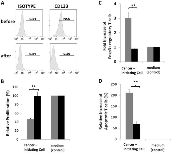
Purpose: Glioblastoma multiforme is a lethal cancer that responds poorly to therapy. Glioblastoma multiforme cancer-initiating cells have been shown to mediate resistance to both chemotherapy and radiation; however, it is unknown to what extent these cells contribute to the profound immunosuppression in glioblastoma multiforme patients and if strategies that alter their differentiation state can reduce this immunosuppression.

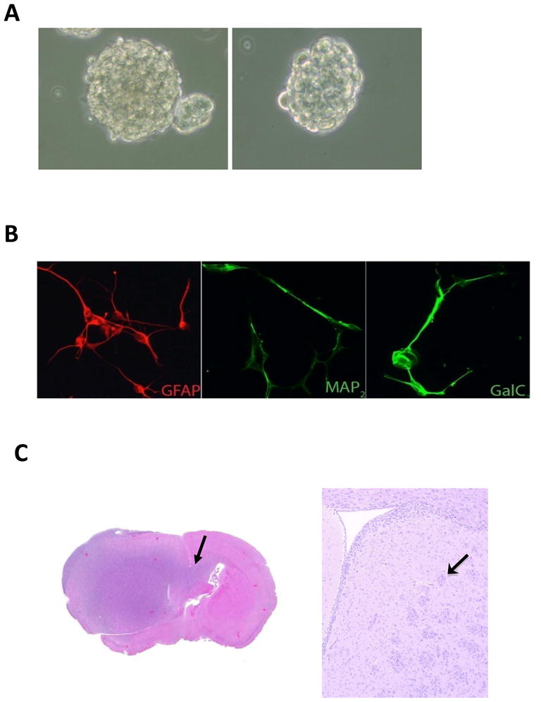
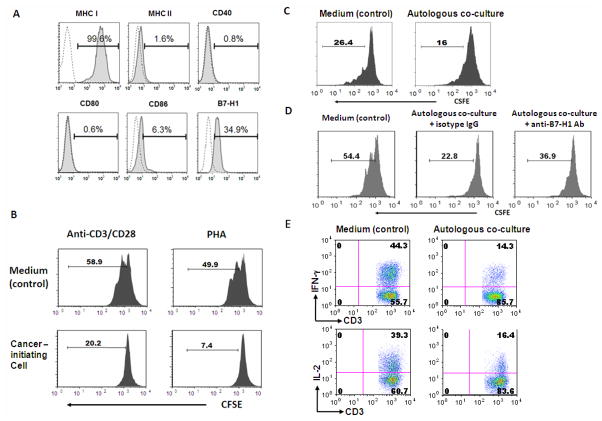
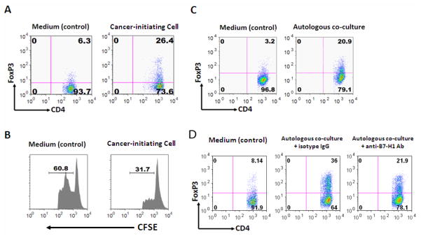
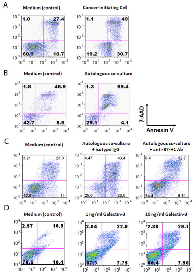
Experimental design: We isolated a subpopulation of cells from glioblastoma multiforme that possessed the capacity for self-renewal, formed neurospheres in vitro, were capable of pluripotent differentiation, and could initiate tumors in vivo. The immune phenotype of these cells was characterized including the elaboration of immunosuppressive cytokines and chemokines by ELISA. Functional immunosuppressive properties were characterized based on the inhibition of T-cell proliferation and effector responses, triggering of T-cell apoptosis, and induction of FoxP3(+) regulatory T cells. On altering their differentiation state, the immunosuppressive phenotype and functional assays were reevaluated.

Results: We found that the cancer-initiating cells markedly inhibited T-cell proliferation and activation, induced regulatory T cells, and triggered T-cell apoptosis that was mediated by B7-H1 and soluble Galectin-3. These immunosuppressive properties were diminished on altering the differentiation of the cancer-initiating cells.

Conclusion: Cancer-initiating cells contribute to tumor evasion of the immunosurveillance and approaches that alter the differentiation state may have immunotherapeutic potential.


Figures

Figure 1
Figure 1. Characterization of human glioma associated cancer-initiating cells from GBM specimens
A. A representative image of neurospheres from one glioma associated cancer-initiating cells is shown. B. After 7 days of culture in differentiating medium, the glioma associated cancer-initiating cells differentiated into GFAP+ astroglial lineage cells, MAP2+ neuronal lineage cells, and GalC+ oligodendroglial lineage cells (magnification × 40) indicating the glioma associated cancer-initiating cells have multi-potent differentiation potential. C. A representative image of a glioma associated cancer-initiating cells xenografted into the frontal lobe of a nude mouse. The tumor that developed from the glioma associated cancer-initiating cells caused enlargement of the brain and were diffusely infiltrative (arrow), including into white matter tracts such as the corpus callosum (arrow).
Figure 2
Figure 2. Glioma associated cancer-initiating cells mediate immunosuppression on human T cells
A. Immune surface phenotype of representative glioma associated cancer-initiating cells. The glioma associated cancer-initiating cells were surface stained with antibodies to MHC I, MHC II, CD40, CD80, CD86, and B7-H1. Representative FACS histogram plots for one glioma associated cancer-initiating cells are shown for target staining (solid line) with associated isotype controls (dotted line). Percentages of the positive populations are shown. B. The glioma associated cancer-initiating cells supernatants inhibit T cell proliferation regardless of activating stimulus. When healthy donor PBMCs were cultured in the presence of the glioma associated cancer-initiating cells supernatants, T cell proliferation was inhibited as demonstrated by FACS analysis of T cell CFSE labeling. Representative FACS histogram plots with the percentages of the indicated cells are shown. C. Glioma associated cancer-initiating cells suppress T cell proliferation through cell-to-cell contact. When autologous PBMCs were cultured in the presence of glioma associated cancer-initiating cells from the same GBM patient, proliferation of T cells was inhibited as demonstrated by FACS analysis with CFSE labeling. A representative FACS histogram plot is shown and similar results were obtained with glioma associated cancer-initiating cells and matched PBMCs from two other patients. Percentages of the indicated populations of proliferated CSFE labeled T cells are shown. D. B7-H1 expressed on glioma associated cancer-initiating cells mediates suppression of T cell proliferation through cell-to-cell contact. When autologous PBMCs were cultured with cancer-initiating cells of the same GBM patient in the presence of B7-H1 neutralizing antibody, the inhibition of T cell proliferation was partially reversed by B7-H1 blockade. The addition of isotype control IgG failed to suppress the inhibition of T cell proliferation, similar to autologous co-culture without IgG as shown in Fig. 2C. E. Glioma associated cancer-initiating cells inhibit T cell function by down-regulating effector cytokine production through cell-to-cell contact. After co-culturing with autologous glioma associated cancer-initiating cells for 3 days, GBM patients’ PBMCs were stimulated with anti-CD3/anti-CD28, surface stained with anti-CD3, and then stained to detect intracellular IFN-γ and IL-2. Data were collected via FACScan. Compared to the medium (control), the glioma associated cancer-initiating cells inhibited both IFN-γ and IL-2 production by gated CD3+ T cells. One representative FACS plot is shown with percentage in upper-right quadrant indicating percentage of positive cells. Similar results were obtained for glioma associated cancer-initiating cells and matched PBMCs from two other patients.
Figure 3
Figure 3. Glioma associated cancer-initiating cells induce functional regulatory T cells
A. The supernatants from the glioma associated cancer-initiating cells induce an increase of the number of FoxP3+ regulatory T cells in the gated CD4+ T cells as shown by the representative FACS analysis. B. FoxP3+ regulatory T cells induced by the supernatants of cancer-initiating cells suppress T cell proliferation. T cells that were treated with cancer-initiating cell supernatants were harvested, co-cultured for 3 days with autologous PBMCs (labeled with CFSE, responder cells) at a 1:1 ratio in the presence of soluble anti-CD3 and subsequently analyzed via FACScan. The number above the line in each histogram represents proliferating responder cells C. Glioma associated cancer-initiating cells also induce FoxP3+ regulatory T cells through cell-to-cell contact. FACS analysis shows that the glioma associated cancer-initiating cells increased the percentage of FoxP3+ regulatory T cells in the gated CD4+ T cells. D. The induction of Foxp3+ regulatory T cells mediated by cell-to-cell contact is partially reduced after the addition of B7-H1 neutralizing antibody. One representative FACS plot is shown with percentage in upper-right quadrant indicating percentage of positive cells. Similar results were obtained for glioma associated cancer-initiating cells and autologous PBMCs from two other patients.
Figure 4
Figure 4. Glioma associated cancer-initiating cells trigger T cell apoptosis
A. After culturing with the glioma associated cancer-initiating cells supernatants, T cells were stimulated with anti-CD3/CD28 and stained with 7-AAD and Annexin V. Compared to medium alone (control), the glioma associated cancer-initiating cells enhanced T cell apoptosis. B. The glioma associated cancer-initiating cells induce T cell apoptosis through cell-to-cell contact. The glioma associated cancer-initiating cells were co-cultured with autologous PBMCs at a 1:10 ratio, and an apoptotic assay was performed after 3 days of culture. C. B7-H1 blockade in cell-to-cell contacting context reduces T cell apoptosis. Both apoptotic and pre-apoptotic T cell percentages decreased when addition of B7-H1 neutralizing antibody was added to culture conditions compared to the isotype control. One representative FACS plot is shown. Similar results were obtained for glioma associated cancer-initiating cells and autologous PBMCs from two other patients. D. Galectin-3 can induce T cell apoptosis in a dose dependent manner within physiological ranges produced by cancer-initiating cells supernatants.
Figure 5
Figure 5. Glioma associated cancer-initiating cells lose immunosuppressive properties upon altering their state of differentiation
Glioma associated cancer-initiating cells were cultured in neural stem cell medium or exposed to differentiating medium for 2–3 passages (5–7 days per passage). A. CD133 expression was reduced on the glioma associated cancer-initiating cells upon exposure to differentiation medium. B. Gioma associated cancer-initiating cells exposed to differentiating medium were less suppressive of T cell proliferation. Healthy donor PBMCs were co-cultured with supernatants from undifferentiated glioma associated cancer-initiating cells or altered differentiated glioma associated cancer-initiating cells in the presence of anti-CD3/CD28 stimulation, and cell proliferation was measured by CCK-8 after 4 days. T cell proliferation in medium alone (control) was used as a baseline (100%), and the relative change in T cell proliferation was plotted relative to this baseline. C. Glioma associated cancer-initiating cells exposed to differentiating medium do not induce FoxP3+ Regulatory T cells. Cultured T cells on day 4 from (A) were stained for CD4 and FoxP3, and FACS data were converted into bar graphs showing fold change in the percentage of FoxP3+ Regulatory T cells versus medium alone (control; set at baseline of 1). The results are averages from three independent experiments, with error bars demonstrating standard deviation. D. Glioma associated cancer-initiating cells exposed to differentiating medium reduces T cell apoptosis. Cultured T cells on day 4 were analyzed for apoptosis, and the increase in apoptotic T cells was calculated as follows: percentage of apoptotic T cells in the presence of supernatants from the glioma associated cancer-initiating cells minus percentage of apoptotic T cells in medium alone (control) divided by percentage of apoptotic T cells in medium alone. In B, C and D, the gray bars represent the results from conditioned medium from glioma associated cancer-initiating cells; black bars represent the results from conditioned medium from differentiated glioma associated cancer-initiating cells. This entire data set was obtained with similar results from two other patients. **P < 0.01 indicates significant difference between undifferentiated and differentiated glioma associated cancer-initiating cells.

Similar articles

Cited by

References

    1. Dingli D, Michor F. Successful therapy must eradicate cancer stem cells. Stem Cells. 2006;24:2603–10. - PubMed
    1. Sanai N, Alvarez-Buylla A, Berger MS. Neural stem cells and the origin of gliomas. N Engl J Med. 2005;353:811–22. - PubMed
    1. Liu G, Yuan X, Zeng Z, et al. Analysis of gene expression and chemoresistance of CD133+ cancer stem cells in glioblastoma. Mol Cancer. 2006;5:67–79. - PMC - PubMed
    1. Sun Y, Kong W, Falk A, et al. CD133 (Prominin) negative human neural stem cells are clonogenic and tripotent. PLoS One. 2009;4:e5498. - PMC - PubMed
    1. Shmelkov SV, Butler JM, Hooper AT, et al. CD133 expression is not restricted to stem cells, and both CD133+ and CD133− metastatic colon cancer cells initiate tumors. J Clin Invest. 2008;118:2111–20. - PMC - PubMed

Publication types

MeSH terms