. 2010 Apr;59(4):1030-7.
doi: 10.2337/db09-1694. Epub 2010 Jan 12.

Inhibition of monocyte adhesion to endothelial cells and attenuation of atherosclerotic lesion by a glucagon-like peptide-1 receptor agonist, exendin-4

Affiliations

Inhibition of monocyte adhesion to endothelial cells and attenuation of atherosclerotic lesion by a glucagon-like peptide-1 receptor agonist, exendin-4

Masayuki Arakawa et al. Diabetes. 2010 Apr.

Abstract

Objective: Exogenous administration of glucagon-like peptide-1 (GLP-1) or GLP-1 receptor agonists such as an exendin-4 has direct beneficial effects on the cardiovascular system. However, their effects on atherosclerogenesis have not been elucidated. The aim of this study was to investigate the effects of GLP-1 on accumulation of monocytes/macrophages on the vascular wall, one of the earliest steps in atherosclerogenesis.

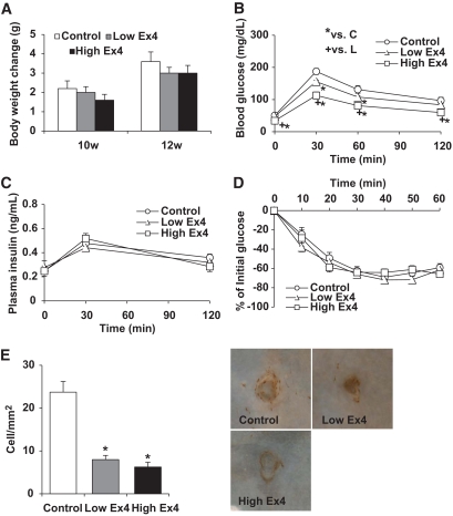
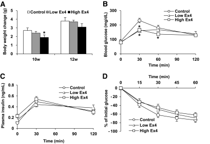
Research design and methods: After continuous infusion of low (300 pmol . kg(-1) . day(-1)) or high (24 nmol . kg(-1) . day(-1)) dose of exendin-4 in C57BL/6 or apolipoprotein E-deficient mice (apoE(-/-)), we evaluated monocyte adhesion to the endothelia of thoracic aorta and arteriosclerotic lesions around the aortic valve. The effects of exendin-4 were investigated in mouse macrophages and human monocytes.

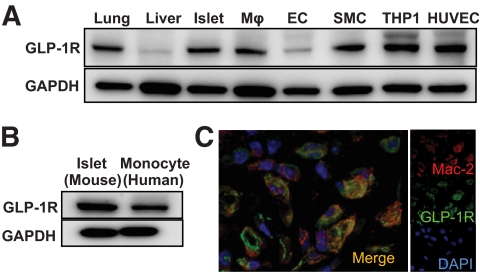
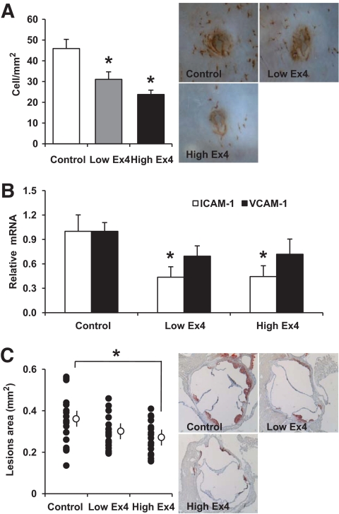
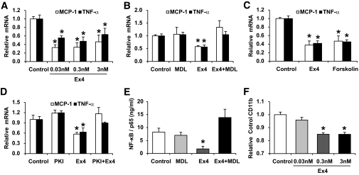
Results: Treatment with exendin-4 significantly inhibited monocytic adhesion in the aortas of C57BL/6 mice without affecting metabolic parameters. In apoE(-/-) mice, the same treatment reduced monocyte adhesion to the endothelium and suppressed atherosclerogenesis. In vitro treatment of mouse macrophages with exendin-4 suppressed lipopolysaccharide-induced mRNA expression of tumor necrosis factor-alpha and monocyte chemoattractant protein-1, and suppressed nuclear translocation of p65, a component of nuclear factor-kappaB. This effect was reversed by either MDL-12330A, a cAMP inhibitor or PKI(14-22), a protein kinase A-specific inhibitor. In human monocytes, exendin-4 reduced the expression of CD11b.

Conclusions: Our data suggested that GLP-1 receptor agonists reduced monocyte/macrophage accumulation in the arterial wall by inhibiting the inflammatory response in macrophages, and that this effect may contribute to the attenuation of atherosclerotic lesion by exendin-4.


Figures

FIG. 1.
FIG. 1.
Expression of GLP-1 receptor on macrophages. A: Expression of GLP-1 receptor in murine lung and liver, isolated murine islets, isolated murine macrophages (Mϕs), cultured murine endothelial cells (ECs), cultured murine smooth muscle cells (SMCs), human monocyte derived line, THP-1 cells, and HUVECs. B: Expression of GLP-1 receptor in human monocytes from healthy subjects. C: Immunohistochemical staining of GLP-1 receptor (green) and Mac-2, a marker of macrophages (red) in atherosclerotic lesions of apoE−/− mice. (A high-quality digital representation of this figure is available in the online issue.)
FIG. 2.
FIG. 2.
Exendin-4 reduced monocytic adhesion to the endothelium in C57BL/6 mice. A: Changes in body weight during treatment with exendin-4 in C57BL/6 mice (n = 6 each). B: Blood glucose concentrations during IPGTT after 24-day treatment with exendin-4 (n = 6 each). C: Plasma insulin levels during IPGTT after 24-day treatment with exendin-4 (n = 6 each). D: Results of insulin tolerance test in each group after 24-day treatment with exendin-4 (n = 6 each). E: The density of adherent Mac-2–positive cells on endothelial cells at branching areas in each group of mice after 28-day treatment (n = 6) with representative en face views of immunohistologic staining with Mac-2 antibody. Data are mean ± SEM. *P < 0.05 versus high-dose group, +P < 0.05 versus low-dose group. (A high-quality digital representation of this figure is available in the online issue.)
FIG. 3.
FIG. 3.
The metabolic effect of exendin-4 in apoE−/− mice. A: Changes in body weight during exendin-4 treatment in apoE−/− mice (n = 13). B: Blood glucose concentrations during IPGTT after 24-day treatment with exendin-4 (n = 6). C: Plasma insulin levels during IPGTT after 24-day treatment with exendin-4 (n = 6). D: Results of insulin tolerance test in each group after 24-day treatment with exendin-4 (n = 6). Data are mean ± SEM. *P < 0.01 versus high-dose group, +P < 0.01 versus low-dose group.
FIG. 4.
FIG. 4.
Exendin-4 reduced monocyte adhesion to the endothelium and atherosclerotic lesions in apoE−/− mice. A: En face immunohistochemical staining with Mac-2 antibody of the aorta of each group. The density of adherent Mac-2–positive cells on the endothelium at branching areas in each group of mice after 28-day treatment (n = 7) and representative en face views of immunohistologic staining with Mac-2 antibody. B: Aortas harvested from each group of mice after 28-day treatment were used for isolation of total RNA. The mRNA expression levels of ICAM-1 and VCAM-1 were determined by quantitative RT-PCR. Relative gene expression is displayed as the level of expression in the test mice relative to that in the control group (set at 1.0, n = 5–7). C: Representative histologic sections of the aortic sinuses stained with oil red O after 28-day treatment. The mean area of oil red O–positive lesions was determined (n = 20). Data are mean ± SEM. *P < 0.05 versus control group. (A high-quality digital representation of this figure is available in the online issue.)
FIG. 5.
FIG. 5.
Exendin-4 reduced the inflammatory response through cAMP signaling pathway in macrophages, and reduced the expression of CD11b in human monocytes. A: Peritoneal macrophages isolated from 8-week-old C57BL/6 mice were incubated with various concentrations of exendin-4 (0.03–3 nmol/l) for 1 h followed by treatment with LPS (1 μg/ml) for 1 h. Then, macrophages were used for isolation of total RNA. The mRNA expression levels of TNF-α and MCP-1 were determined by quantitative RT-PCR. Relative gene expression is displayed as the level of expression in peritoneal macrophages without the addition of exendin-4 set at 1.0 (n = 4–5). B: Peritoneal macrophages were preincubated with 5 μmol/l MDL-12330A for 30 min before the addition of 0.3 nmol/l exendin-4 and then incubated with LPS (1 μg/ml) for 1 h. Then, macrophages were used for isolation of total RNA (n = 4–6). C: Peritoneal macrophages were incubated with 0.3 nmol/l exendin-4 or 10 μmol/l forskolin for 1 h followed by LPS (1 μg/ml) for 1 h. Then, macrophages were used for isolation of total RNA (n = 4–5). D: Peritoneal macrophages were preincubated with 10 μmol/l PKI14-22 for 30 min before the addition of 0.3 nmol/l exendin-4 and then incubated with LPS (1 μg/ml) for 1 h. Then, macrophages were used for isolation of total RNA (n = 4–5). E: Peritoneal macrophages were preincubated with 5 μmol/l MDL-12330A for 30 min before the addition of 0.3 nmol/l exendin-4 and then incubated with LPS (1 μg/ml) for 1 h. Then, macrophages were used for isolation of nuclear protein extracts. The nuclear level of NF-κB p65 was determined by enzyme-linked immunosorbent assay (ELISA) (n = 3–4). F: Human monocytes isolated from healthy volunteers were incubated without or with various concentrations of exendin-4 (0.03–3 nmol/l) for 24 h. Then, the surface expression of CD11b was assessed by flow cytometry. Data are median fluorescence intensity relative to the control. *P < 0.05 versus the control group.

References

    1. Greig NH, Holloway HW, De Ore KA, Jani D, Wang Y, Zhou J, Garant MJ, Egan JM: Once daily injection of exendin-4 to diabetic mice achieves long-term beneficial effects on blood glucose concentrations. Diabetologia 1999; 42: 45– 50 - PubMed
    1. Kolterman OG, Buse JB, Fineman MS, Gaines E, Heintz S, Bicsak TA, Taylor K, Kim D, Aisporna M, Wang Y, Baron AD: Synthetic exendin-4 (exenatide) significantly reduces postprandial and fasting plasma glucose in subjects with type 2 diabetes. J Clin Endocrinol Metab 2003; 88: 3082– 3089 - PubMed
    1. Parkes DG, Pittner R, Jodka C, Smith P, Young A: Insulinotropic actions of exendin-4 and glucagon-like peptide-1 in vivo and in vitro. Metabolism 2001; 50: 583– 589 - PubMed
    1. Dupré J, Behme MT, McDonald TJ: Exendin-4 normalized postcibal glycemic excursions in type 1 diabetes. J Clin Endocrinol Metab 2004; 89: 3469– 3473 - PubMed
    1. Szayna M, Doyle ME, Betkey JA, Holloway HW, Spencer RG, Greig NH, Egan JM: Exendin-4 decelerates food intake, weight gain, and fat deposition in Zucker rats. Endocrinology 2000; 141: 1936– 1941 - PubMed

MeSH terms