. 2010 Jan 15;70(2):532-42.
doi: 10.1158/0008-5472.CAN-09-1477. Epub 2010 Jan 12.

Transcriptional autoregulation by BRCA1

Affiliations

Transcriptional autoregulation by BRCA1

Adriana De Siervi et al. Cancer Res. .

Abstract

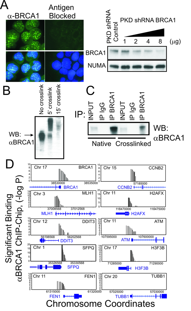
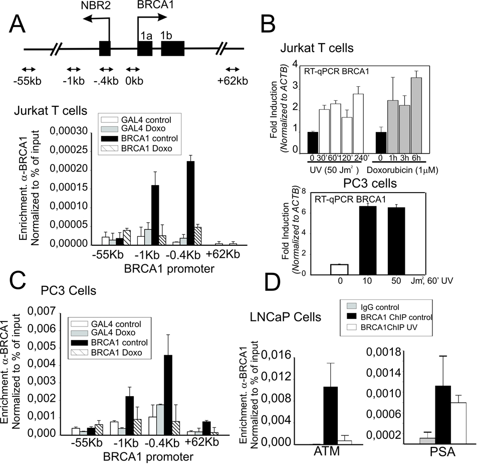
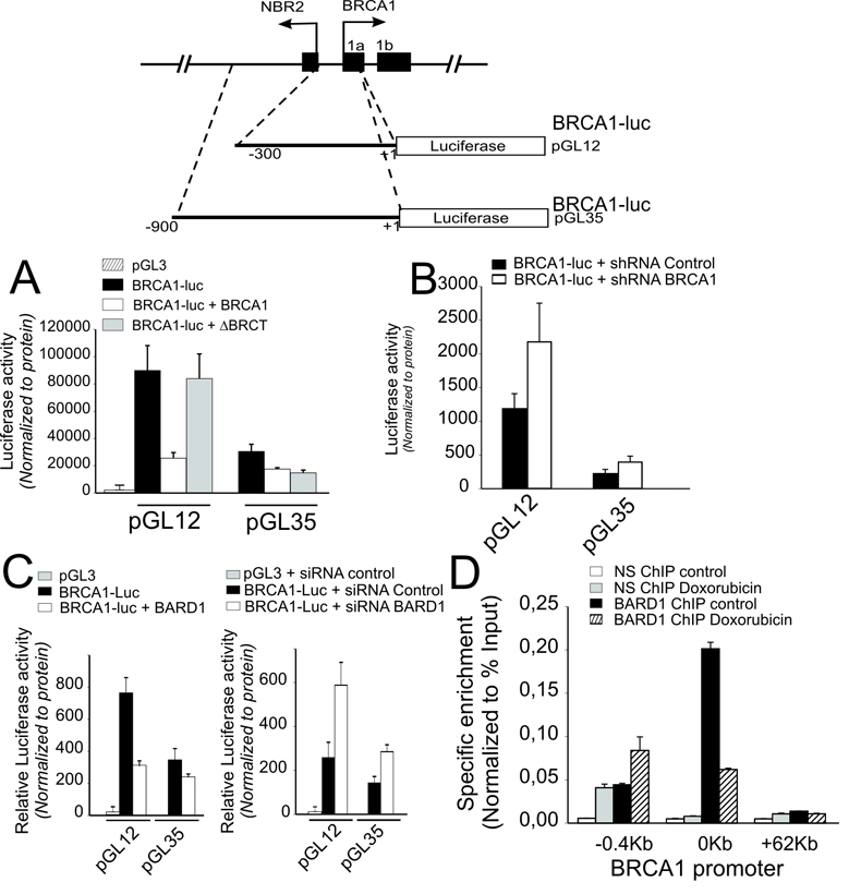
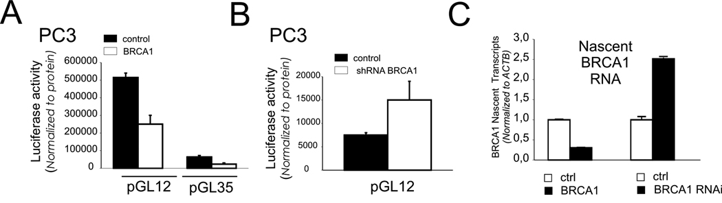
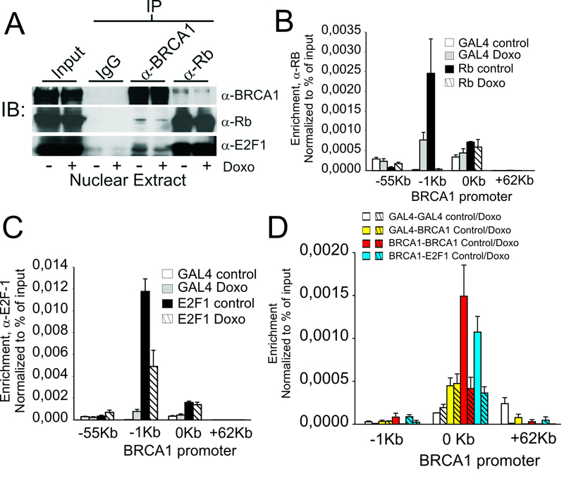
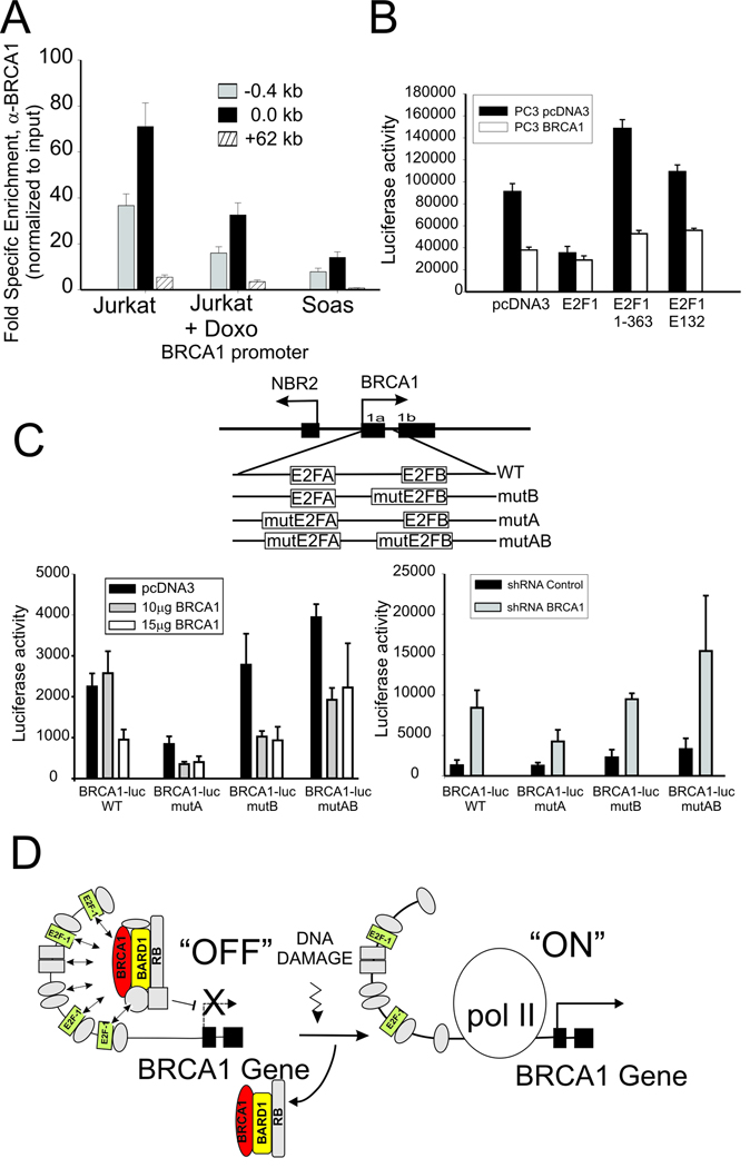
The BRCA1 gene product plays numerous roles in regulating genome integrity. Its role in assembling supermolecular complexes in response to DNA damage has been extensively studied; however, much less is understood about its role as a transcriptional coregulator. Loss or mutation is associated with hereditary breast and ovarian cancers, whereas altered expression occurs frequently in sporadic forms of breast cancer, suggesting that the control of BRCA1 transcription might be important to tumorigenesis. Here, we provide evidence of a striking linkage between the roles for BRCA1 as a transcriptional coregulator with control of its expression via an autoregulatory transcriptional loop. BRCA1 assembles with complexes containing E2F-1 and RB to form a repressive multicomponent transcriptional complex that inhibits BRCA1 promoter transcription. This complex is disrupted by genotoxic stress, resulting in the displacement of BRCA1 protein from the BRCA1 promoter and subsequent upregulation of BRCA1 transcription. Cells depleted of BRCA1 respond by upregulating BRCA1 transcripts, whereas cells overexpressing BRCA1 respond by downregulating BRCA1 transcripts. Tandem chromatin immmunoprecipitation studies show that BRCA1 is regulated by a dynamic coregulatory complex containing BRCA1, E2F1, and Rb at the BRCA1 promoter that is disrupted by DNA-damaging agents to increase its transcription. These results define a novel transcriptional mechanism of autoregulated homeostasis of BRCA1 that selectively titrates its levels to maintain genome integrity in response to genotoxic insult.


Figures

Figure 1
Figure 1. BRCA1 protein associates with the promoter regions of BRCA1 and multiple other genes
(A) Characterization of affinity purified antibodies against BRCA1. (left) by Immunofluorescence staining of Jurkat T-cells before and after antigen blockade. α-BRCA1: Green; DAPI DNA staining: Blue (right). Characterization of α-BRCA1 antibodies by immunoblot analysis of cells lysates from Jurkat T-cells depleted of BRCA1 by transfection with control and increasing amounts of BRCA1 shRNA producing plasmid. NuMA antibody was used as loading control. (B) Immunoblot characterization of α-BRCA1 antibody reactivity against formalin cross-linked BRCA1 protein in Jurkat cell lysates. (C) Characterization of α-BRCA1 antibodies by immunoprecipitation of native and formalin cross-linked cell lysates compared to non-specific IgG. Following immunoprecipitation, formalin cross-links were reversed prior to SDS PAGE and immunoblot analysis. Input indicates signal from 10% of lysate. (D) BRCA1 ChIP profile of BRCA1 and other genes that show significant association (p ≤0.000001) with BRCA1 by ChIP-chip analysis using the Nimblegen HG17 tiled proximal promoter array (HG17 array 2005-04-18_min_promoter_set) compared to non-specific control (TUBB1). Results are the average of two independent biological replicates. Y-axis is shown in −log (P) scale and X-axis shows coordinates of the indicated chromosomes.
Figure 2
Figure 2. BRCA1 protein association with the BRCA1 promoter and BRCA1 expression is regulated by genotoxic stress
(A) Inset above, schematic diagram of BRCA1 promoter show divergent NBR2 gene and relative positions of primers set used in this study. ChIP analysis at the BRCA1 promoter using BRCA1-specific or non-specific control IgG (α-Gal4) in untreated Jurkat T-cells or cells treated 3h with doxorubicin (1µM). Shown is enrichment at positions of the BRCA1 locus relative to the transcription start site presented as percent recovery of input. (B) Jurkat T cells or PC3 cells were treated with either UV or doxorubicin (1µM) and harvested at the indicated times prior to RNA isolation. Mature RNA message was determined by RT-qPCR normalized to ACTB. (C) ChIP analysis of BRCA1 binding to the BRCA1 promoter in PC3 human prostate cell lines at indicated positions relative to the TSS in untreated and Doxorubin treated cells (1µM, 24 h). IgG against yeast Gal4 α-Gal4 was used as a non-specific binding control. (D) ChIP analysis of BRCA1 binding to the ATM and PSA promoters in LNCaP cells untreated or after UV treatment. In all cases (1A, C and D) mean enrichment and error shown correspond to the average and standard deviation of two independent experiments.
Figure 3
Figure 3. BRCA1 and BARD1 negatively regulate transcription from the BRCA1 promoter
(A) Jurkat T cells were cotransfected with luciferase reporter constructs (see schematic above) spanning the indicated lengths of regulatory sequences upstream of the BRCA1 TSS (pGL12 or pGL35) in combination with either: BRCA1 wt (pcDNA3 BRCA1), BRCA1 C-terminal mutant (pcDNA3 ΔBRCT) or empty (pcDNA3) expression vectors. (B) Jurkat T-cells were cotransfected with the indicated BRCA1 promoter reporters and either shRNA BRCA1 or control shRNA with scrambled sequence expression vectors. (C) Jurkat T-cells were cotransfected with the indicated BRCA1 promoter reporters and either wild type BARD1 (BARD1) or empty expression vectors or BARD1 siRNA or control duplex siRNA oligonucleotides. Error bars show the standard error of the mean from at least 3 independent transfections. (D) ChIP analysis of BARD1 enrichment at the BRCA1 promoter with and without pretreatment with 1 µM doxorubicin. Error bars indicate the standard error of the mean derived from 2 independent biological replicates.
Figure 4
Figure 4. Reciprocal regulation of BRCA1 promoter activation and transcription in PC3 prostate cell lines by stable over-expression or depletion of BRCA1 protein
(A) PC3 cells stably transfected with control empty vector (ctrl) or BRCA1 expression vector (BRCA1) were transiently transfected with BRCA1-luc reporter constructs (pGL12 or pGL35). (B) PC3 cells stably expressing shRNA BRCA1 or control shRNA with scrambled sequence shRNA expression vectors were transfected with BRCA1-luc reporter (pGL12). Luciferase activity was normalized to protein. Error bars show the standard error of the mean from 3 independent transfections. (C) PC3 prostate cancer cells that stably over-expressing BRCA1 as in (A) were analyzed for nascent BRCA1 RNA levels (unspliced transcript) by qRT-PCR and compared to cells stably expressing empty vector (ctrl). Similarly, cells stably depleted of BRCA1 as in (B) were analyzed for nascent BRCA1 RNA levels compared to cells stably producing shRNA containing a scrambled sequence (ctrl).
Figure 5
Figure 5. BRCA1 forms a negative regulatory complex with E2F-1 and Rb at the BRCA1 promoter that is disrupted by genotoxic stress
(A) Co-immuno-precipitation of BRCA, Rb and E2F-1 using antibodies against non-specific IgG, α-Rb and α-BRCA1 antibodies. (B) ChIP-qPCR analysis of Rb enrichment at the indicated positions of the BRCA1 promoter region in untreated Jurkat T cells and cells treated 3 h with 1 µM doxorubicin compared to non-specific control (α-Gal4). (C) α-E2F-1 ChIP-qPCR analysis of E2F-1 enrichment at the indicated positions of the BRCA1 promoter region in untreated Jurkat T cells and cells treated 3 h with 1 µM doxorubicin compared to non-specific control (α-Gal4). (D) Untreated Jurkat cells or cells treated with 1 µM doxorubicin were analyzed by tandem BRCA1/E2F-1 ChIP using combinations of first ChIP with either non-specific antibody (α-Gal4), or α-BRCA1 antibody followed by Re-ChIP of the immunoprecipitated complexes with α-Gal4, α-BRCA1 or α-E2F-1. Mean enrichment and error shown correspond to the average and standard deviation of the mean of at least two independent experiments.
Figure 6
Figure 6. BRCA1 assembles at the BRCA1 promoter in association with E2F-1 and Rb complexes
(A) BRCA1 enrichment by ChIP at BRCA1 promoter of untreated and Doxorubicin treated Jurkat cells compared to untreated Rb-deficient Saos cells normalized to nonspecific IgG. Error bar indicates standard error and mean from 2 independent experiments. (B) PC3 prostate cancer cells stably over-expressing BRCA1 (PC3 BRCA1) or empty vector (PC3 pcDNA3) were co-transfected with the BRCA1-luc reporter plasmid (pGL12) and wild type or the indicated mutant E2F-1 expression vectors (see text). Error bars show the standard error of the mean from 3 independent transfections. (C) BRCA1 regulation of the BRCA1 promoter depends on the E2F binding sites. BRCA1 was over-expressed (left) or depleted (right) in cells transfected with reporter plasmid containing wild type or mutated E2F transcription factor binding sites within the BRCA1 promoter sequences (see schematic diagram above). Result are the mean and standard error from 3 independent transfections. (D) Hypothetical schematic diagram depicting BRCA1 negative autoregulation by a complex containing BRCA1, E2F-1 and Rb at the BRCA1 promoter, which is up-regulated by displacement/disruption of this complex at the promoter to allow BRCA1 transcription in response to genotoxic stress. Note that E2F-1 complexes are rearranged but not completely lost from the promoter.

Similar articles

Cited by

References

    1. Miki Y, Swensen J, Shattuck-Eidens D, et al. A strong candidate for the breast and ovarian cancer susceptibility gene BRCA1. Science. 1994;266:66–71. - PubMed
    1. Shen SX, Weaver Z, Xu X, et al. A targeted disruption of the murine Brca1 gene causes gamma-irradiation hypersensitivity and genetic instability. Oncogene. 1998;17:3115–3124. - PubMed
    1. Deng CX. BRCA1: cell cycle checkpoint, genetic instability, DNA damage response and cancer evolution. Nucleic Acids Res. 2006;34:1416–1426. - PMC - PubMed
    1. Rosen EM, Fan S, Ma Y. BRCA1 regulation of transcription. Cancer Lett. 2006;236:175–185. - PubMed
    1. Mullan PB, Quinn JE, Harkin DP. The role of BRCA1 in transcriptional regulation and cell cycle control. Oncogene. 2006;25:5854–5863. - PubMed

Publication types