. 2010 Jan 15;70(2):481-9.
doi: 10.1158/0008-5472.CAN-09-1688. Epub 2010 Jan 12.

Natural killer cell cytotoxicity is suppressed by exposure to the human NKG2D ligand MICA*008 that is shed by tumor cells in exosomes

Affiliations

Natural killer cell cytotoxicity is suppressed by exposure to the human NKG2D ligand MICA*008 that is shed by tumor cells in exosomes

Omodele Ashiru et al. Cancer Res. .

Abstract

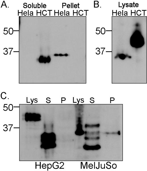
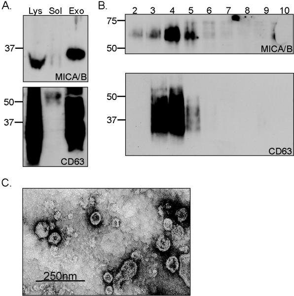
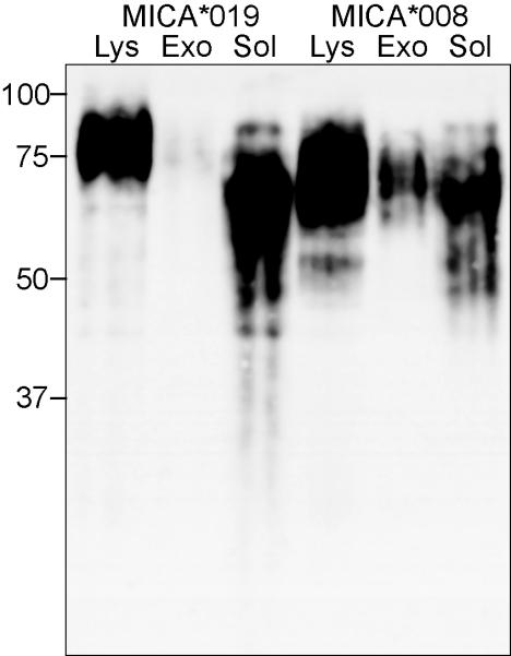
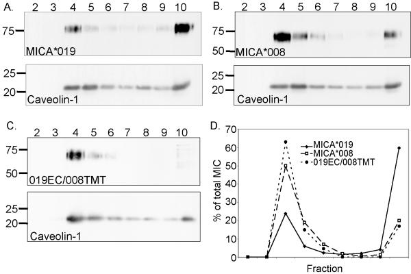
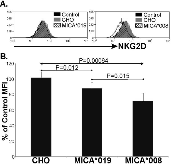
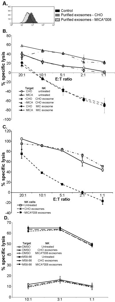
The MHC class I-related chain (MIC) A and MICB ligands for the activating receptor NKG2D can be shed from tumor cells, and the presence of these soluble molecules in sera is related with compromised immune response and progression of disease. Recently, thiol disulphide isomerases and members of the ADAM (a disintegrin and metalloproteinase) gene family were identified as key enzymes in mediating MICA/B shedding from cells. Here, we report shedding of the most frequently expressed MICA allele in human populations (MICA*008) into exosomes, small membrane vesicles that are secreted upon fusion with the plasma membrane. Although similar to other MICA/B molecules in the extracellular domain, the predicted transmembrane and cytoplasmic domains of MICA*008 are quite different, and this difference seemed to be critical for the mode of release from tumor cells. Treatment of natural killer (NK) cells with exosomes containing MICA*008 molecules not only triggered downregulation of NKG2D from the cell surface but also provoked a marked reduction in NK cytotoxicity that is independent of NKG2D ligand expression by the target cell. Our findings reveal a mechanism of NK suppression in cancer that may facilitate immune escape and progression.


Figures

Figure 1
Figure 1. MICA molecules released from Hela pellet at 100,000 xg
(A.) Supernatants were collected from cells that had been cultured for 16 hours centrifuged to eliminate large membrane fragments and organelles and then fractionated to separate proteins that pellet at 100,000 xg, from soluble proteins (recovered by TCA precipitation). Samples were digested with PNGase F before western blot analysis. (B.) Total cell lysates were analysed for comparison. (C.) Analysis of MICA/B released by the tumour cell lines HepG2 and MelJuSo. Lys – lysate, S – soluble, P – pellet. Data are representative of three experiments. Positions of Mr weight markers (kDa) are indicated.
Figure 2
Figure 2. Exosome preparations from Hela contain MICA and are enriched in the tetraspanin CD63
A. Cells were cultured for 16 hours in serum free medium and exosomes were isolated. The fractions corresponding to soluble protein (sol), exosomes (exo) and total cell lysate (lys) were analysed by western blot, probing with either anti-MICA antibody or anti-CD63, as indicated. Samples were digested with PNGase F before western blot analysis for MICA. B. Exosomes were further fractionated on a step sucrose gradient and fractions analysed for MICA and CD63. C. Exosomes were visualised by electron microscopy. Data are representative of two experiments.
Figure 3
Figure 3. MICA*008, but not MICA*019, is released in exosomes
Exosomes and soluble proteins were isolated from cell free culture supernatants of CHO cells stably transfected with either MICA*019 or MICA*008. The preparations of exosomes and soluble proteins were analysed by western blot (3 experiments).
Figure 4
Figure 4. The majority of MICA*008 is present in detergent resistant membranes (DRMs)
CHO cells transfected with MICA*019 (A), MICA*008 (B), or a chimaeric protein comprising the extracellular domain of MICA*019 fused to the transmembrane and cytoplasmic tail of MICA*008 (019EC/008TMT) (C) were lysed and fractionated by centrifugation on sucrose gradients. Equal volumes of these fractions were analysed, by western blot, for the presence of MICA. Data are representative of at least three experiments. (D) Quantitative analysis to determine the proportion of MICA protein present in each fraction was carried out using Image J software.
Figure 5
Figure 5. Both soluble and exosomal MICA downmodulate surface expression of NKG2D
Culture supernatants were collected from CHO cells transfected with MICA*019 or *008. Supernatant from untransfected CHO cells was used as control. A. Supernatant from CHO cells and MICA transfected CHO cells were incubated with IL-2 activated human NK cells for 24 hours. The amount of NKG2D on the surface of the NK cells was then analysed by flow cytometry. The result shown is representative of 5 experiments. B. Downmodulation of NKG2D is significant (Student’s T Test) after treatment with either MICA*019 or *008 containing supernatants. Data are expressed as a percentage of the NKG2D expression observed on NK cells incubated in medium alone.
Figure 6
Figure 6. Exosomal MICA*008 can trigger both NKG2D downmodulation and compromise NK cell cytotoxicity
Incubation of NK cells with purified exosomes isolated from CHO cells transfected with MICA*008, but not untransfected CHO cells, leads to a marked reduction in NKG2D cell surface expression (A) (4 experiments) and compromised NK cell mediated lysis of parental and MICA transfected CHO cells (B), as well as unrelated “third-party” cells such as 721.221 (C). Note that AlamarBlue measures cell metabolism, thus negative values for percentage specific lysis indicate target cell proliferation. Pre-incubation of CTL specific for the influenza matrix peptide M58-66 bound to HLA-A2, with either control or MICA*08 containing exosomes has no effect on specific cytotoxicity (D) (2 experiments).

References

    1. Bauer S, Groh V, Wu J, et al. Activation of NK cells and T cells by NKG2D, a receptor for stress- inducible MICA. Science. 1999;285:727–9. - PubMed
    1. Maasho K, Opoku-Anane J, Marusina AI, Coligan JE, Borrego F. NKG2D is a costimulatory receptor for human naive CD8+ T cells. J Immunol. 2005;174:4480–4. - PubMed
    1. Verneris MR, Karami M, Baker J, Jayaswal A, Negrin RS. Role of NKG2D signaling in the cytotoxicity of activated and expanded CD8+ T cells. Blood. 2004;103:3065–72. - PubMed
    1. Gonzalez S, Groh V, Spies T. Immunobiology of human NKG2D and its ligands. Curr Top Microbiol Immunol. 2006;298:121–38. - PubMed
    1. Groh V, Bahram S, Bauer S, Herman A, Beauchamp M, Spies T. Cell stress-regulated human major histocompatibility complex class I gene expressed in gastrointestinal epithelium. Proc Natl Acad Sci U S A. 1996;93:12445–50. - PMC - PubMed

Publication types

MeSH terms

Substances