

Matrix metalloproteinase-9 functions as a tumor suppressor in colitis-associated cancer
- PMID: 20068187
- PMCID: PMC2821688
- DOI: 10.1158/0008-5472.CAN-09-3166
Matrix metalloproteinase-9 functions as a tumor suppressor in colitis-associated cancer
Abstract
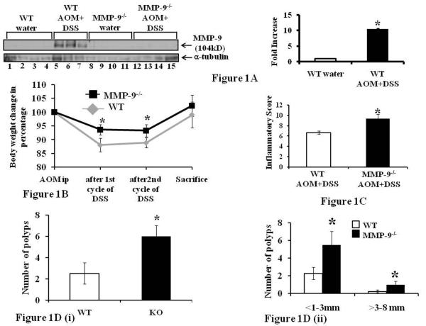
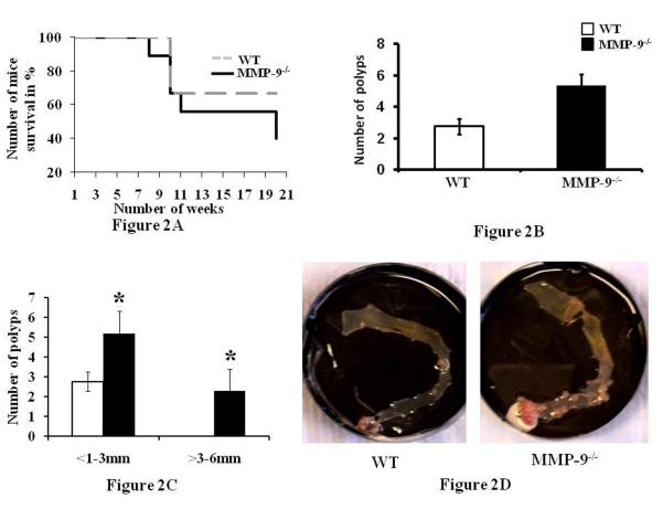
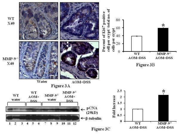
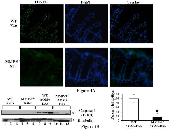
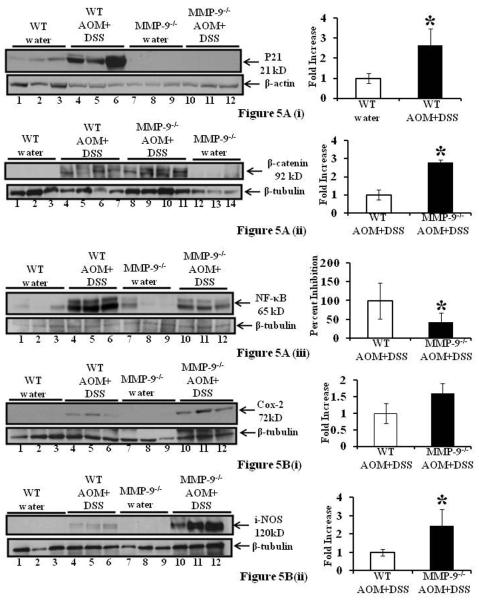
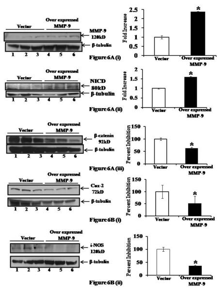
There is a well-documented association of matrix metalloproteinase-9 (MMP-9) and receptor Notch-1 overexpression in colon cancer. We recently showed that MMP-9 is also upregulated in colitis, where it modulates tissue damage and goblet cell differentiation via proteolytic cleavage of Notch-1. In this study, we investigated whether MMP-9 is critical for colitis-associated colon cancer (CAC). Mice that are wild type (WT) or MMP-9 nullizygous (MMP-9(-/-)) were used for in vivo studies and the human enterocyte cell line Caco2-BBE was used for in vitro studies. CAC was induced in mice using an established carcinogenesis protocol that involves exposure to azoxymethane followed by treatment with dextran sodium sulfate. MMP-9(-/-) mice exhibited increased susceptibility to CAC relative to WT mice. Elevations in tumor multiplicity, size, and mortality were associated with increased proliferation and decreased apoptosis. Tumors formed in MMP-9(-/-) mice exhibited expression of p21(WAF1/Cip1) and increased expression of beta-catenin relative to WT mice. In vitro studies of MMP-9 overexpression showed increased Notch-1 activation with a reciprocal decrease in beta-catenin. Notch and beta-catenin/Wnt signaling have crucial roles in determining differentiation and carcinogenesis in gut epithelia. Despite being a mediator of proinflammatory responses in colitis, MMP-9 plays a protective role and acts as a tumor suppressor in CAC by modulating Notch-1 activation, thereby resulting in activation of p21(WAF1/Cip1) and suppression of beta-catenin.
Figures
Similar articles
-
Epithelial derived-matrix metalloproteinase (MMP9) exhibits a novel defensive role of tumor suppressor in colitis associated cancer by activating MMP9-Notch1-ARF-p53 axis.Oncotarget. 2017 Jan 3;8(1):364-378. doi: 10.18632/oncotarget.13406. Oncotarget. 2017. PMID: 27861153 Free PMC article.
-
Notch1 regulates the effects of matrix metalloproteinase-9 on colitis-associated cancer in mice.Gastroenterology. 2011 Oct;141(4):1381-92. doi: 10.1053/j.gastro.2011.06.056. Epub 2011 Jun 30. Gastroenterology. 2011. PMID: 21723221 Free PMC article.
-
Upregulated claudin-1 expression promotes colitis-associated cancer by promoting β-catenin phosphorylation and activation in Notch/p-AKT-dependent manner.Oncogene. 2019 Jun;38(26):5321-5337. doi: 10.1038/s41388-019-0795-5. Epub 2019 Apr 10. Oncogene. 2019. PMID: 30971761 Free PMC article.
-
Matrix metalloproteinase 9 (MMP9) limits reactive oxygen species (ROS) accumulation and DNA damage in colitis-associated cancer.Cell Death Dis. 2020 Sep 17;11(9):767. doi: 10.1038/s41419-020-02959-z. Cell Death Dis. 2020. PMID: 32943603 Free PMC article.
-
Matrix metalloproteinase-9 regulates MUC-2 expression through its effect on goblet cell differentiation.Gastroenterology. 2007 May;132(5):1877-89. doi: 10.1053/j.gastro.2007.02.048. Epub 2007 Feb 23. Gastroenterology. 2007. PMID: 17484881
Cited by
-
Fluctuating roles of matrix metalloproteinase-9 in oral squamous cell carcinoma.ScientificWorldJournal. 2013;2013:920595. doi: 10.1155/2013/920595. Epub 2013 Jan 8. ScientificWorldJournal. 2013. PMID: 23365550 Free PMC article. Review.
-
Casticin Inhibits A375.S2 Human Melanoma Cell Migration/Invasion through Downregulating NF-κB and Matrix Metalloproteinase-2 and -1.Molecules. 2016 Mar 19;21(3):384. doi: 10.3390/molecules21030384. Molecules. 2016. PMID: 27007357 Free PMC article.
-
The immunomodulatory role of matrix metalloproteinases in colitis-associated cancer.Front Immunol. 2023 Jan 19;13:1093990. doi: 10.3389/fimmu.2022.1093990. eCollection 2022. Front Immunol. 2023. PMID: 36776395 Free PMC article. Review.
-
Inflammation-Associated Carcinogenesis in Inflammatory Bowel Disease: Clinical Features and Molecular Mechanisms.Cells. 2025 Apr 9;14(8):567. doi: 10.3390/cells14080567. Cells. 2025. PMID: 40277893 Free PMC article. Review.
-
Epithelial derived-matrix metalloproteinase (MMP9) exhibits a novel defensive role of tumor suppressor in colitis associated cancer by activating MMP9-Notch1-ARF-p53 axis.Oncotarget. 2017 Jan 3;8(1):364-378. doi: 10.18632/oncotarget.13406. Oncotarget. 2017. PMID: 27861153 Free PMC article.
References
-
- Egeblad M, Werb Z. New functions for the matrix metalloproteinases in cancer progression. Nat Rev Cancer. 2002;2:161–74. - PubMed
-
- Okamoto R, Watanabe M. Cellular and molecular mechanisms of the epithelial repair in IBD. Dig Dis Sci. 2005;50(Suppl 1):S34–8. - PubMed
-
- Castaneda FE, Walia B, Vijay-Kumar M, et al. Targeted deletion of metalloproteinase 9 attenuates experimental colitis in mice: central role of epithelial-derived MMP. Gastroenterology. 2005;129:1991–2008. - PubMed
-
- Baugh MD, Perry MJ, Hollander AP, et al. Matrix metalloproteinase levels are elevated in inflammatory bowel disease. Gastroenterology. 1999;117:814–22. - PubMed
Publication types
MeSH terms
Substances
Grants and funding
LinkOut - more resources
Full Text Sources
Molecular Biology Databases
Miscellaneous
