. 2010 May;18(5):1026-34.
doi: 10.1038/mt.2009.315. Epub 2010 Jan 12.

Mesenchymal stem cells expressing osteogenic and angiogenic factors synergistically enhance bone formation in a mouse model of segmental bone defect

Affiliations

Mesenchymal stem cells expressing osteogenic and angiogenic factors synergistically enhance bone formation in a mouse model of segmental bone defect

Sanjay Kumar et al. Mol Ther. 2010 May.

Abstract

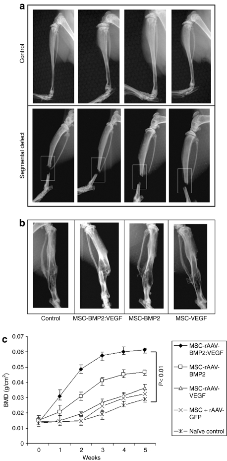
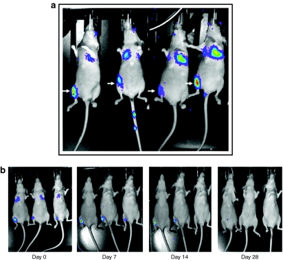
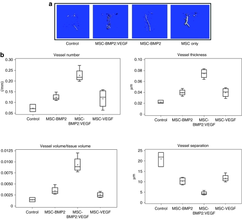
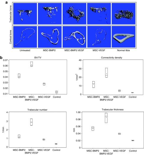
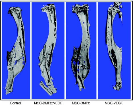
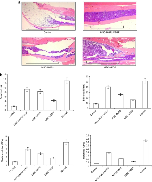
The potential of mesenchymal stem cells (MSC) in tissue regeneration is increasingly gaining attention. There is now accumulating evidence that MSC make an important contribution to postnatal vasculogenesis. During bone development and fracture healing, vascularization is observed before bone formation. The present study determined the potential of MSC, transduced ex vivo with a recombinant adeno-associated virus 6 (rAAV6) encoding bone morphogenetic protein 2 (BMP2) and vascular endothelial growth factor (VEGF) in a mouse model of segmental bone defect created in the tibiae of athymic nude mice. Mouse MSC that were mock-transduced or transduced with rAAV6-BMP2:VEGF were systemically transplanted following radiographic confirmation of the osteotomy. Effects of the therapy were determined by enzyme-linked immunosorbent assay measurements for BMP2 and VEGF, dual-energy X-ray absorptiometry (DXA) for bone density, three-dimensional microcomputed tomography (microCT) for bone and capillary architecture, and histomorphometry for bone remodeling. Results of these analyses indicated enhanced bone formation in the group that received BMP2+VEGF-expressing MSC compared to other groups. The therapeutic effects were accompanied by increased vascularity and osteoblastogenesis, indicating its potential for effective use while treating difficult nonunion bone defects in humans.


Figures

<b>Figure 1</b>
Figure 1
Bone mineral density (BMD) in the segmental defect area of tibiae during fracture healing. (a) Segmental defect was surgically created in the right tibia of 10–12-week-old nude mice. (b) X-ray imaging of mice after 5 weeks of MSC transplantation showing the fracture healing process. (c) A total of 1 × 10⁶ MSC that were unmodified, transduced with rAAV6-BMP2:VEGF or rAAV6-GFP, were administered in five consecutive days by intravenous injection. Mice were anesthetized with isoflurane for dual-energy X-ray absorptiometry (DXA) analysis. BMD was determined weekly around the fractured area of tibia by noninvasive DXA to follow the bone growth. DXA was performed in a GE Lunar PIXImus machine, and data analyses were performed using PIXImus software version 1.43.020. BMP2, bone morphogenetic protein 2; MSC, mesenchymal stem cells; rAAV, recombinant adeno-associated virus; VEGF, vascular endothelial growth factor.
<b>Figure 2</b>
Figure 2
Biodistribution of systemically transplanted luciferase-positive MSC. (a) Luciferase-positive MSCs were intravenously transplanted into nude mice, following creation of tibial segmental defect in the right leg, and bioluminescence image was monitored after 24 hours. Representative images of mice showing homing-in-on of transplanted MSC to the site of segmental defect are shown above. (b) Bioluminescence imaging was performed at indicated time points to track long-term viability and existence of transplanted, luciferase-positive MSC in recipient mice. MSC, mesenchymal stem cells.
<b>Figure 3</b>
Figure 3
MSC-expressing osteogenic and angiogenic factors show vascularized bone development around segmental defect area of tibia and microarchitectural parameters of Microfil-perfused vasculature of tibia. (a) After intravenous transplantation of MSC, mice were sacrificed after Microfil perfusion from each group, and bones were used for µCT. Representative µCT images of vasculature in Microfil-perfused tibia (around the segmental defect area) show three-dimensional image of region of interest (ROI) extracted from reconstructed vasculature volume after 5 weeks of treatment of mice in control group; those injected with MSC-expressing BMP2 and VEGF, those injected with MSC-expressing BMP2, and those injected with MSC-expressing VEGF are shown. (b) Denoted values of microarchitectural parameters were determined from three-dimensional µCT measurements from groups of mice receiving AAV-BMP2:VEGF treatment or no treatment. Horizontal lines in each box from top to bottom indicate 25th, 50th, and 75th percentiles. Error bars indicate 10th and 90th percentiles. AAV, adeno-associated virus; BMP2, bone morphogenetic protein 2; µCT, microcomputed tomography; MSC, mesenchymal stem cells; VEGF, vascular endothelial growth factor.
<b>Figure 4</b>
Figure 4
µCT analysis of cortical and trabecular bones of fractured tibia and microarchitectural parameters of trabecular bone around the fractured tibia after MSC therapy. (a) Tibia from MSC-transplanted cohorts of mice were used for µCT analysis of cortical bone and trabecular bones. Representative images from indicated groups show three-dimensional images of tibia extracted from reconstructed bone volume, 8 weeks after treatments. (b) Denoted values of microarchitectural parameters were determined from three-dimensional µCT measurements from groups of mice receiving indicated treatments or no treatment (control). Horizontal lines in each box from top to bottom indicate 25th, 50th, and 75th percentiles. Error bars indicate 10th and 90th percentiles. BMP2, bone morphogenetic protein 2; µCT, microcomputed tomography; MSC, mesenchymal stem cells; VEGF, vascular endothelial growth factor.
<b>Figure 5</b>
Figure 5
µCT analysis of fixed tibia after MSC therapy. Tibiae from MSC-transplanted cohorts of mice were used for µCT analysis. Representative images from indicated groups show three-dimensional images of tibiae extracted from reconstructed bone volume, 16 weeks after the treatment. BMP2, bone morphogenetic protein 2; µCT, microcomputed tomography; MSC, mesenchymal stem cells; VEGF, vascular endothelial growth factor.
<b>Figure 6</b>
Figure 6
Osteogenic and angiogenic factors induce synergistic enhancement of bone healing in a segmental defect of tibia. (a) Histology of bone sections after H&E staining indicates significant increase of newly formed bone in the area of osteotomy, following treatment with MSC-expressing BMP2 and VEGF, compared to other groups. The region of segmental defect is indicated by horizontal lines in each panel. (b) Three-point bending tests (peak load, stiffness) and nanoindentation (elastic modulus and hardness) were performed on tibia isolated from the MSC-transplanted and control mice after 16 weeks of treatment. Data shown represent mean ± SD. *P < 0.05. BMP2, bone morphogenetic protein 2; H&E, hematoxylin and eosin; MSC, mesenchymal stem cells; VEGF, vascular endothelial growth factor.

References

    1. Praemer A, Furner S., and , Rice DP. Musculoskeletal Conditions in the United States. American Academy of Orthopaedic Surgeons: Rosemont; 1999.
    1. Nolta JA.ed.) (2006Gene Therapy for Mesenchymal Stem Cells. Springer Science
    1. Franceschi RT. Biological approaches to bone regeneration by gene therapy. J Dent Res. 2005;84:1093–1103. - PubMed
    1. Ylä-Herttuala S., and , Alitalo K. Gene transfer as a tool to induce therapeutic vascular growth. Nat Med. 2003;9:694–701. - PubMed
    1. Ferrara N., and , Gerber HP. The role of vascular endothelial growth factor in angiogenesis. Acta Haematol. 2001;106:148–156. - PubMed

Publication types

Substances

LinkOut - more resources