. 2010 May;18(5):1035-45.
doi: 10.1038/mt.2009.293. Epub 2010 Jan 12.

Dendritic cells transduced with lentiviral vectors expressing VIP differentiate into VIP-secreting tolerogenic-like DCs

Affiliations

Dendritic cells transduced with lentiviral vectors expressing VIP differentiate into VIP-secreting tolerogenic-like DCs

Miguel G Toscano et al. Mol Ther. 2010 May.

Abstract

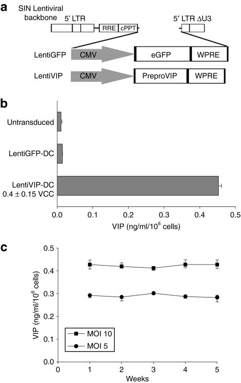
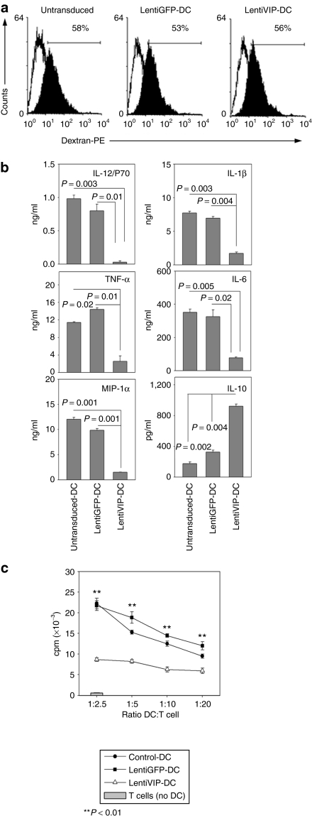
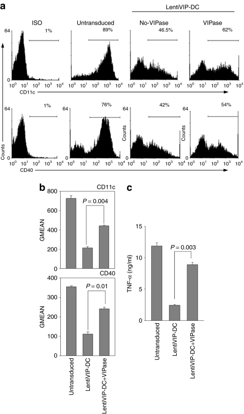
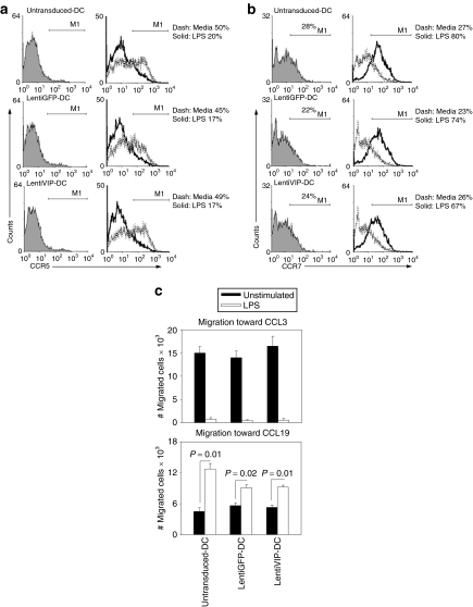
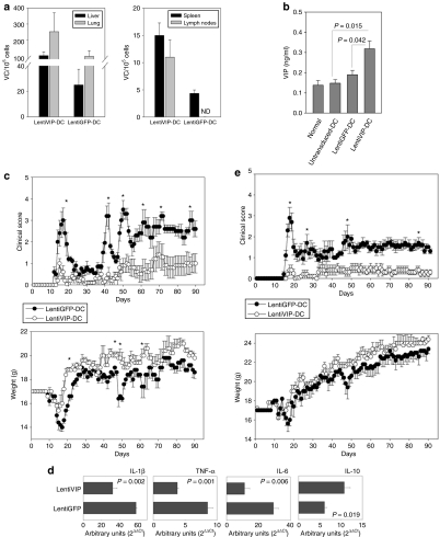
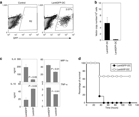
Dendritic cells (DCs) initiate immune responses as well as tolerance. We showed previously that the neuropeptide vasoactive intestinal peptide (VIP) suppresses innate immune responses, modulates adaptive responses by generating regulatory T cells (Treg) through the induction of tolerogenic DCs (tDCs), and has therapeutic effects in models of autoimmune/inflammatory disorders. Systemic VIP administration is limited by its short biological half-life and by its pleiotropic effects on the cardiovascular system and gastrointestinal tract. Therefore, we used lentiviral vectors to genetically engineer VIP-expressing bone marrow-derived DC (BMDC) and characterized the transduced LentiVIP-DC in terms of phenotype and therapeutic effects in models of experimental autoimmune encephalomyelitis (EAE) and cecal ligation and puncture (CLP) sepsis. LentiVIP-DCs secrete VIP, and resemble tDCs through lack of co-stimulatory molecule upregulation, lack of proinflammatory cytokine secretion, increased interleukin (IL)-10 production, and poor stimulation of allogeneic T cells. A single inoculation of LentiVIP-DC in EAE or CLP mice had therapeutic effects, which correlated with reduced expression of proinflammatory cytokines and increased IL-10 production in spinal cord and peritoneal fluid, respectively. In contrast to systemic VIP administration that requires repeated, high-dose inoculations, local delivery of VIP by LentiVIP-DC may represent a promising therapeutic tool for the treatment of autoimmune diseases and inflammatory disorders.


Figures

<b>Figure 1</b>
Figure 1
Bone marrow–derived dendritic cells are efficiently transduced with lentiviral vectors expressing vasoactive intestinal peptide (VIP) and green fluorescent protein (GFP). (a) Schematic representation of lentiviral vectors LentiVIP and LentiGFP. Cytomegalovirus (CMV) promoter drives the expression of preproVIP complementary DNA in LentiVIP and of GFP in LentiGFP in a self-inactivating lentiviral vector backbone. (b) VIP secretion by LentiVIP-DC. LentiVIP-DC were cultured for 24 hours at a concentration of 1 × 10⁶ cells/ml. Supernatants were collected and assayed for VIP by competitive enzyme-linked immunosorbent assay. Undetectable levels of VIP in untransduced- and LentiGFP-DC. (c) Stable secretion of VIP over time by LentiVIP-DC at multiplicity of infection (MOI) 5 and 10. Once a week, cells were washed, resuspended in fresh medium, supernatants were collected 24 hours later and VIP levels were determined as described in b. One representative experiment out of three is shown. cPPT, polypurine track; LTR, long terminal repeat; RRE, Rev responsive element; WPRE, woodchuck postregulatory element.
<b>Figure 2</b>
Figure 2
Phenotypic characterization of LentiVIP-DC. (a) Untransduced-DC, LentiGFP-DC, and LentiVIP-DC harvested on day 7 were washed and stained with antibodies to CD11c, I-Ek, CD40, CD80, and CD86 and analyzed by flow cytometry. (b) Dendritic cells were treated with 1 µg/ml lipopolysaccharide (LPS) for 24 hours. Histograms are representative of three experiments performed in duplicate. (c) Results of fluorescence-activated cell-sorting analysis expressed as geometric mean of fluorescence (GMEAN) ± SD of three experiments performed in duplicate. Degree of significance P < 0.05 compared with untransduced-DC and LentiGFP-DC. Final GMEAN values are the result of GMEAN subtraction from isotype control. (d) LentiVIP-DC are CD11low and CD45RBhigh. Untransduced-DC and LentiVIP-DC were double stained for CD11c and CD45RB expression and analyzed by flow cytometry. CD11clowCD45RBhigh population is gated as R3. One representative experiment of three is shown.
<b>Figure 3</b>
Figure 3
Functional characteristics of LentiVIP-DC. (a) Untransduced-, LentiGFP-, and LentiVIP-DC were incubated in medium containing 1 mg/ml of dextran-PE (40 kd) for 2 hours at 4 °C (control; open histograms) or 37 °C (filled histograms), extensively washed and analyzed by flow cytometry. One representative experiment of two performed in duplicate is shown. (b) Untransduced-, LentiGFP-, and LentiVIP-DC were cultured with LPS (1 µg/ml) for 24 hours. Culture supernatants were assayed for IL-12p70, IL-1β, TNF-α, IL-6, MIP-1α, and IL-10. Results are the mean ± SD of three experiments performed in duplicate. Degree of significance P < 0.05. (c) Untransduced-, LentiGFP-, and LentiVIP-DC were cocultured at different ratios with allogeneic CD4+ T cells (5 × 10⁴) in a 4-day mixed lymphocyte reaction. Proliferation was measured by [3H]-thymidine incorporation. A representative experiment of three is shown.
<b>Figure 4</b>
Figure 4
VIPase catalytic antibody partially reverses the characteristics of LentiVIP-DC. LentiVIP-DC were grown in the presence of daily added 180 µg of VIPase or control antibody (UPC10 IgG2a) as described in Materials and Methods. (a) LentiVIP-DC treated with or without VIPase were stained with anti-CD11c, or treated for 24 hours with lipopolysaccharide (LPS) and stained for CD40 expression. (b) Results are expressed as geometric mean of fluorescence (GMEAN) ± SD of three experiments performed in duplicate. Degree of significance P < 0.05. (c) TNF-α levels were determined by enzyme-linked immunosorbent assay in supernatants of LPS-treated cells. Three experiments were performed in duplicate. Degree of significance P < 0.05.
<b>Figure 5</b>
Figure 5
CCR5 and CCR7 patterns in LentiVIP-DC. (a) CCR5 and (b) CCR7 expression was analyzed by flow cytometry in untransduced-, LentiGFP-, and LentiVIP-DC before and after lipopolysaccharide (LPS) activation. Cells were treated with LPS (0.1 µg/ml) for 24 hours before detection of CCR5 and 48 hours before detection of CCR7. Indirect immunostaining was performed using a primary purified CCR5 or 7 antibody, a secondary biotin-conjugated antibody, and APC-conjugated streptavidin. Filled: isotype. Dashed line: not activated. Solid line: LPS-activated. (c) Migration toward CCL3 (100 ng/ml) and CCL19 (100 ng/ml) was tested for untransduced-, LentiVIP-, and LentiGFP-DC, before and after LPS treatment. A volume of 2 × 10⁵ cells were plated and allowed to migrate for 4 hours. Absolute number of cells determined by FACS (120-second counts) are presented. Number of migrated cells in control wells without chemokines were <1,000 cells in all of the cases (data not shown). Two independent experiments in triplicate were performed. Degree of significance P < 0.05.
<b>Figure 6</b>
Figure 6
LentiVIP-DC reduces severity in two experimental autoimmune encephalomyelitis (EAE) models. (a) Liver, lung, spleen, and lymph nodes (axillary and inguinal) were collected 48 hours after intravenous inoculation of transduced cells and genomic DNA was isolated. The presence of LentiVIP-DC and LentiGFP-DC was analyzed by quantitative PCR using primers specific for sequences for each vector. Presented data are after subtraction of background from tissues of control mice. (b) Serum from LentiVIP-DC, LentiGFP-DC, and untransduced-DC SJL-treated mice was obtained 48 hours after the dendritic cell (DC) inoculation and vasoactive intestinal peptide (VIP) levels were measured by enzyme-linked immunosorbent assay and compared with endogenous levels of VIP in serum of healthy mice. (c) SJL/J mice (5 mice/group) immunized with PLP139–151 as described in Methods were inoculated intravenous (tail vein) on day 6 with LentiVIP-DC or LentiGFP-DC (3 × 10⁶ cells) pulsed with 50 µg/ml of PLP (3 × 10⁶). Clinical score and weight was followed daily for >90 days. (d) Total RNA was extracted from spinal cord of LentiVIP-DC and LentiGFP-DC SJL-treated animals at the peak of disease and analyzed for cytokine expression by real-time qPCR. The expression of cytokines was normalized to GAPDH and is relative to healthy controls (no EAE). Degree of significance P < 0.05. (e) C57BL/6 mice (6 mice/group) were immunized with MOG35–55 as described in Materials and Methods. On day 8, the mice were inoculated intravenous with 3 × 10⁶ LentiGFP-DC or LentiVIP-DC pulsed with 50 µg/ml of MOG. Clinical score and weight was followed daily for >90 days. *P < 0.05. IL, interleukin; TNF, tumor necrosis factor.
<b>Figure 7</b>
Figure 7
LentiVIP-DC increases survival rate in the cecal ligation and puncture model. Male BALB/c mice (6–8 weeks old; 6/group) were subjected to cecal ligation and puncture as described in Materials and Methods. Immediately after the surgical procedure LentiVIP-DC and LentiGFP-DC (2 × 10⁶ cells/animal) were injected intraperitoneal and 20 hours after the surgical procedure peritoneal exudate cells and fluid were collected. (a) LentiGFP-DC cells were detected by flow cytometry in the peritoneal cavity. (b) LentiVIP was detected by quantitative PCR after DNA extraction from peritoneal exudate cells. Primers to detect LentiVIP were used; LentiGFP-DC was used as negative control. (c) Peritoneal fluid was analyzed by enzyme-linked immunosorbent assay for cytokine production. Degree of significance P < 0.05. (d) The therapeutic effect of LentiVIP-DC was evaluated by survival rate over time. GFP, green fluorescent protein; IL, interleukin; MIP, macrophage inflammatory protein; TNF, tumor necrosis factor.

Similar articles

Cited by

References

    1. Wolfraim LA. Treating autoimmune diseases through restoration of antigen-specific immune tolerance. Arch Immunol Ther Exp (Warsz) 2006;54:1–13. - PubMed
    1. Roth JC, Curiel DT., and , Pereboeva L. Cell vehicle targeting strategies. Gene Ther. 2008;15:716–729. - PubMed
    1. Steinman RM. The dendritic cell system and its role in immunogenicity. Annu Rev Immunol. 1991;9:271–296. - PubMed
    1. Banchereau J., and , Steinman RM. Dendritic cells and the control of immunity. Nature. 1998;392:245–252. - PubMed
    1. Steptoe RJ., and , Thomson AW. Dendritic cells and tolerance induction. Clin Exp Immunol. 1996;105:397–402. - PMC - PubMed

Publication types

MeSH terms

Substances