. 2010 Apr;18(4):743-53.
doi: 10.1038/mt.2009.304. Epub 2010 Jan 12.

Zinc-finger nuclease-induced gene repair with oligodeoxynucleotides: wanted and unwanted target locus modifications

Affiliations

Zinc-finger nuclease-induced gene repair with oligodeoxynucleotides: wanted and unwanted target locus modifications

Sarah Radecke et al. Mol Ther. 2010 Apr.

Abstract

Correcting a mutated gene directly at its endogenous locus represents an alternative to gene therapy protocols based on viral vectors with their risk of insertional mutagenesis. When solely a single-stranded oligodeoxynucleotide (ssODN) is used as a repair matrix, the efficiency of the targeted gene correction is low. However, as shown with the homing endonuclease I-SceI, ssODN-mediated gene correction can be enhanced by concomitantly inducing a DNA double-strand break (DSB) close to the mutation. Because I-SceI is hardly adjustable to cut at any desired position in the human genome, here, customizable zinc-finger nucleases (ZFNs) were used to stimulate ssODN-mediated repair of a mutated single-copy reporter locus stably integrated into human embryonic kidney-293 cells. The ZFNs induced faithful gene repair at a frequency of 0.16%. Six times more often, ZFN-induced DSBs were found to be modified by unfaithful addition of ssODN between the termini and about 60 times more often by nonhomologous end joining-related deletions and insertions. Additionally, ZFN off-target activity based on binding mismatch sites at the locus of interest was detected in in vitro cleavage assays and also in chromosomal DNA isolated from treated cells. Therefore, the specificity of ZFN-induced ssODN-mediated gene repair needs to be improved, especially regarding clinical applications.


Figures

<b>Figure 1</b>
Figure 1
System outline: target locus of the 293/3-1 cells and the repair matrices for DSB repair. An integrated single copy of the plasmid pCMV.LacZs31δGFP (7,832 bp) constitutes the target locus. CMV IE: cytomegalovirus immediate early promoter/enhancer. LacZ: complete ORF encoding β-galactosidase. δEGFP: EGFP ORF with a 5′-deletion of 32 bp rendering EGFP nonfunctional. pA: SV40 polyadenylation signal. Gray wavy lines symbolize the flanking chromosomal sequences. The nucleotide sequence of the locus depicts the region where the 3′-terminus of the LacZ ORF is joined via a 42-bp fragment to the 5′-deleted EGFP ORF. For translational decoupling, stop codons (underlined) are introduced in frames +0 and +1. The binding sites for I-SceI and the ZFNs are highlighted with the color-coded jagged arrows pointing toward the respective cut sites. An AseI recognition site forms the 6-bp spacer region. ssODNs (all of sense orientation): primary structures of the repair and control matrices. Note that for 3-1corr, one version without and one with a BdT were available. 3-1c-s-BdT has a 5′-terminal 40-bp region homologous to the intact LacZ ORF whereas the remainder of the molecule—being identical to the 3′ part of 3-1scr—is unable to reconstitute the EGFP ORF. 3-1scr is composed of the nucleotides of 3-1corr but in a scrambled order. dsDNA matrix: the nucleotide sequence of this positive control covers the target site where both reporter ORFs are to be fused in frame. It has 2,697 repair-relevant base pairs: 5′ homology region of 1,950 bp (δLacZ ORF, 1,950 bp); repair segment of 41 bp (9-bp-stuffer; 32 bp reconstructing the 5′-terminus of EGFP ORF); 3′ homology region of 706 bp (688 bp specific for EGFP ORF, plus additional 18 bp abutting the EGFP ORF). Ovals mark the start codon of the cut with EGFP ORF. Scheme of pUC.Zgfp (5,845 bp): This plasmid harbors the dsDNA matrix. Note that this control was always used in its linearized form (E: EcoRI) with dephosphorylated 5′ termini to reduce direct circularization upon cell entry. Further sequence informations about the dsDNA matrix can be found in Supplementary Figure S7. Drawings are not to scale. BdT, biotinylated thymidine; bp, base pair; DSB, double-strand break; dsDNA, double-stranded DNA; EGFP, enhanced green fluorescent protein; ORF, open reading frame; ssODN, single-stranded oligodeoxynucleotide; ZFN, zinc-finger nuclease.
<b>Figure 2</b>
Figure 2
Chromosomal repair rates. 293-3/1 cells were analyzed 7 days post-transfection. EGFP+ cells among the living (7-AAD) cell population were counted by FACS. Mimicking the situation of an ex vivo protocol where corrected cells cannot necessarily be enriched from noncorrected ones, repair rates were related to all living cells, i.e., no normalization was calculated. Transfection quality controls at 5 hours postnucleofection (see Supplementary Materials and Methods) resulted in nine valid independent experiments the data of which are presented in box plot format. Black bar: median; right edge of the gray box: 75% quartile; left edge of the box: 25% quartile; the horizontal line connects the minimum value with the maximum value. MOCK: nuclease-negative control with plasmid pRK5, the backbone-only version of the expression plasmids. In the mock samples for the matrices, cells received a calcium phosphate-precipitate with no DNA added. Statistical significance (P = 0.05) was tested with the two-tailed Wilcoxon matched pair signed-rank test: Null hypothesis not rejected (*), rejected (**, ***). dsDNA, double-stranded DNA; EGFP, enhanced green fluorescent protein; FACS, fluorescence-activated cell sorting.
<b>Figure 3</b>
Figure 3
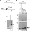
Capture assay analyzing the fate of an ssODN during targeted gene correction. The 292-bp fragment (gray arrow head) amplified with primers gfpM and gfp7 from the internal control IC17 monitored the functionality of the PCR and the completeness of the NaOH-mediated bottom strand removal. (a) Schemes of the biotinylated test fragment TF (top) and of a biotinylated target locus-specific gDNA fragment (bottom). TF served as locus-specific positive control for the capturing step also allowing the sensitivity of the PCR-based detection step to be assessed. TF was generated by hybridizing 3-1corr-BdT to the bottom strand of a PCR product (primer pair: #48-LacZ-TC/pabox4; template: corrected locus) followed by a one-round elongation. The 452-bp product arises from TF based on its bottom strand. Drawings are not to scale. (b) PCR results of capture assays with gDNAs from sorted EGFP and EGFP+ cells. To separate the PCR products in 1.5% agarose gels, 10% of the reaction volumes were loaded per lane. Data from two independent experiments are presented (input: 200 cells (I), 500 cells (II)). The indicated percentages for IC and TF compare their molecule numbers with the numbers of the target alleles (which are equivalent to the number of cells). (c) Results of the capture assay probing whether the biotinylated ssODN is fully ligated into the top strand. From experiment II, a second sample of 500 EGFP+ cells transfected with 3-1corr-BdT was split into two aliquots one of which was NaOH treated before the PCR. M: 100-bp DNA Ladder. Numbers next to the lanes M indicate fragment lengths in bp. The gels were stained with ethidium bromide and are shown in reverse contrast. bp, base pair; EGFP, enhanced green fluorescent protein; gDNA, genomic DNA; IC, internal control; ssODN, single-stranded oligodeoxynucleotide; TF, test fragment.
<b>Figure 4</b>
Figure 4
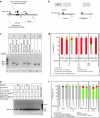
Analyses of unwanted events at the targeted site in EGFP− cells. (a–d) Capture assay based analyses. (a) Scheme of a target locus-specific gDNA fragment carrying an integrated part of a biotinylated ssODN at the DSB site. Black line: top strand; gray line: bottom strand. Primers #48-LacZ-TC and gfp7 encompass the repair site resulting in fragments of different lengths depending on the respective DSB rejoining. The original target locus yields a 465-bp long fragment. The primer pair gfp1/gfp7 amplifies a region of the EGFP ORF 42-bp downstream of the GZF1-N binding site assessing the total number of captured biotinylated alleles (see also Supplementary Figure S4). (b) Scheme depicting the effect of the NaOH treatment on the PCR amplification when the targeted loci contain a 3′-ligated ssODN. All drawings are not to scale. (c) PCR result of capture assays (primer pair #48-LacZ-TC/gfp7). For each sample, 250 ng of gDNA (corresponding to an estimated number of ~28,000 targeted alleles) were utilized. TF monitored the removal of the bottom strand. Ten percentage of the PCR products were analyzed. (d) Summary of sequence results from c. The numbers on top of the columns refer to the total numbers (σ) of sequences which were scored as independently generated. (e–f) Biotin-independent analyses. (e) Gel-electrophoretic separation of products directly amplified from targeted loci. For each sample, 100 ng (~11,100 target alleles) were employed. Digestions of the gDNAs were carried out with AseI to reduce background generated from unmodified target loci. DraI (also cutting an AT-rich hexamer sequence) was used to control the AseI-specific enrichment of modified loci, and to monitor the ensuing PCR with primer pair #48-LacZ-TC/gfp7 in the presence of high numbers of gDNA fragments. (f) Summary of the sequence results from samples of e. The numbers on top of the columns refer to the total numbers (σ) of sequences which were scored as independently generated. All gel pictures are presented in reverse contrast. M: 100-bp DNA ladder. Numbers next to the marker lane M indicate fragment lengths in bp. bp, base pair; DSB, double-strand break; EGFP, enhanced green fluorescent protein; gDNA, genomic DNA; ORF, open reading frame; ssODN, single-stranded oligodeoxynucleotide.
<b>Figure 5</b>
Figure 5
In vitro assay of zinc-finger nuclease activity. (a) Digestion of the circular target locus plasmid pCMV.LacZs31δGFP by in vitro-made ZFNs. Reaction products were separated in a 0.8% agarose gel and stained with ethidium bromide. The picture is shown in reverse contrast. As positive control for linearization (lane 11), pCMV.LacZs31δGFP was digested with BamHI. L: 1-kb DNA ladder. Numbers to the right of the marker lane L indicate fragment lengths in kb. (b) Scheme of the artificial linear substrate with both 5′-DIG top and 5′-DIG bottom label. Numbers indicate the lengths (nts) of the expected single-stranded digestion products. Canonical binding sites are uniformly highlighted and the ZFN name is written with standard letters. Nonoptimal binding sites are named with italic letters, matches are highlighted. The second GZF1-N-specific site (nts 292–300 of the EGFP ORF) has been located by in silico analyses. The jagged triangles point to the cleavage positions. The gray thick lines refer to the respective spacers with boxed numbers indicating their lengths in bp. Drawing is not to scale. (c) Digestion of a linear DIG-labeled substrate. The reaction products rendered single-stranded were separated in a 1.5% agarose gel and transferred onto membranes. Labeled DNA strands were visualized with a DIG-specific antibody coupled to alkaline phosphatase. 5′-DIG top, 5′-DIG bottom, 5′-DIG top + bottom: PCR products carrying a DIG-label either in the top strand, the bottom strand, or in both. Numbers to the left indicate the lengths (nts) of the original DNA strands (black arrowhead) and the corresponding products (gray arrowheads). Note that these unspecific digestion products in lanes 4, 8, 9, 12, and 13 were observed in 3 out of a total of 5 independent experiments. The reason for this is unknown. bp, base pair; DIG, digoxigenin; nt, nucleotide; ZFN, zinc-finger nuclease.
<b>Figure 6</b>
Figure 6
Recapitulating gene repair experiment outcomes. The boxes represent the different categories of alleles found in the cell population. The categories are listed in descending order according to their frequencies. “>>“: many more alleles as compared to the following category. Note that not all alleles can be detected due to failures in, e.g., bead-capturing, PCR amplification, subcloning. Thus, the rankings are not based on absolute numbers of the different alleles detected, but describe the findings semiquantitatively. DSB, double-strand break.

Similar articles

Cited by

References

    1. Cavazzana-Calvo M, Hacein-Bey S, de Saint Basile G, Gross F, Yvon E, Nusbaum P, et al. Gene therapy of human severe combined immunodeficiency (SCID)-X1 disease. Science. 2000;288:669–672. - PubMed
    1. Gaspar HB, Parsley KL, Howe S, King D, Gilmour KC, Sinclair J, et al. Gene therapy of X-linked severe combined immunodeficiency by use of a pseudotyped gammaretroviral vector. Lancet. 2004;364:2181–2187. - PubMed
    1. Hacein-Bey-Abina S, Von Kalle C, Schmidt M, McCormack MP, Wulffraat N, Leboulch P, et al. LMO2-associated clonal T cell proliferation in two patients after gene therapy for SCID-X1. Science. 2003;302:415–419. - PubMed
    1. Howe SJ, Mansour MR, Schwarzwaelder K, Bartholomae C, Hubank M, Kempski H, et al. Insertional mutagenesis combined with acquired somatic mutations causes leukemogenesis following gene therapy of SCID-X1 patients. J Clin Invest. 2008;118:3143–3150. - PMC - PubMed
    1. Hacein-Bey-Abina S, Garrigue A, Wang GP, Soulier J, Lim A, Morillon E, et al. Insertional oncogenesis in 4 patients after retrovirus-mediated gene therapy of SCID-X1. J Clin Invest. 2008;118:3132–3142. - PMC - PubMed

Publication types

Substances

LinkOut - more resources