. 2010 Feb 2;102(3):594-601.
doi: 10.1038/sj.bjc.6605495. Epub 2010 Jan 12.

A novel fragment derived from the beta chain of human fibrinogen, beta43-63, is a potent inhibitor of activated endothelial cells in vitro and in vivo

Affiliations

A novel fragment derived from the beta chain of human fibrinogen, beta43-63, is a potent inhibitor of activated endothelial cells in vitro and in vivo

E Krajewska et al. Br J Cancer. .

Abstract

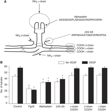
Background: Angiogenesis and haemostasis are closely linked within tumours with many haemostatic proteins regulating tumour angiogenesis. Indeed we previously identified a fragment of human fibrinogen, fibrinogen E-fragment (FgnE) with potent anti-angiogenic properties in vitro and cytotoxic effects on tumour vessels in vivo. We therefore investigated which region of FgnE was mediating vessel cytotoxicity.

Methods: Human dermal microvascular endothelial cells (ECs) were used to test the efficacy of peptides derived from FgnE on proliferation, migration, differentiation, apoptosis and adhesion before testing the efficacy of an active peptide on tumour vasculature in vivo.

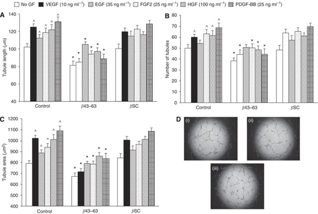
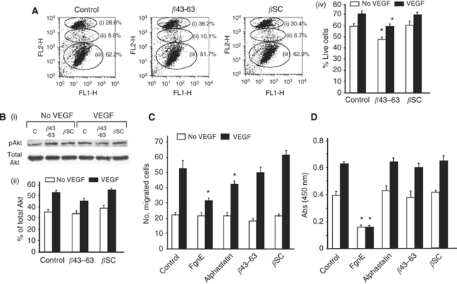
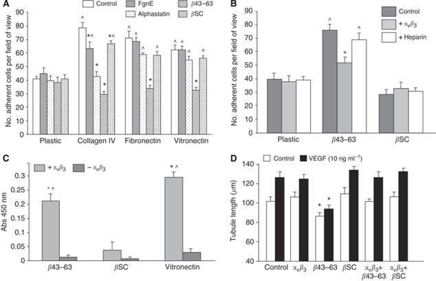
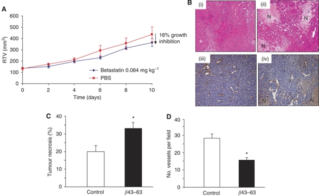
Results: We identified a 20-amino-acid peptide derived from the beta chain of FgnE, beta43-63, which had no effect on EC proliferation or migration but markedly inhibited the ability of activated ECs to form tubules or to adhere to various constituents of the extracellular matrix - collagen IV, fibronectin and vitronectin. Furthermore, our data show that beta43-63 interacts with ECs, in part, by binding to alpha(v)beta(3), so soluble alpha(v)beta(3) abrogated beta43-63 inhibition of tubule formation by activated ECs. Finally, when injected into mice bearing tumour xenografts, beta43-63 inhibited tumour vascularisation and induced formation of significant tumour necrosis.

Conclusions: Taken together, these data suggest that beta43-63 is a novel anti-tumour peptide whose anti-angiogenic effects are mediated by alpha(v)beta(3).


Figures

Figure 1
Figure 1
(A) Schematic illustration of FgnE. Fibrinogen consists of two α chains, two β chains and two γ chains. Alphastatin is the first 24 amino acids of the N terminus of the α chains (α1–24) whereas β43–63 is the first 20 amino acids on the N terminus of the two β chains. 20-amino-acid peptides were also generated from the C terminus of each chain representing the regions exposed upon plasmin cleavage of the original fibrinogen molecule. (B) Effects of FgnE and derived peptides on the Matrigel assay. FgnE, alphastatin and β43–63 inhibit tubule formation in response to VEGF stimulation as measured by number of tubules per field of view. FgnE was used at 1 μM, but the peptides were used at 2 μM as two copies of these peptides are present in a single FgnE molecule. Data are presented as mean±s.e.m. *P<0.05 with respect to relevant control.
Figure 2
Figure 2
β43–63 also inhibits tubule formation by ECs in response to four other pro-angiogenic growth factors, PDGF, FGF2, EGF and HGF, in vitro. (A) Length, (B) number and (C) area covered by tubules formed by HuDMECs in response to the above growth factors in the absence or presence of β43–63 or scrambled β43–63 (βSC) used at 2 μM. *P<0.05 with respect to relevant control group, ^P<0.05 with respect to control No-GF group. All data are means±s.e.m. Data are representative of three replicate experiments. (D) Typical appearance of VEGF-induced tubules (in wells of a 96-well plate) in the presence of (i) VEGF alone, (ii) VEGF + 2 μM β43–63 and (iii) VEGF + 2 μM βSC.
Figure 3
Figure 3
Effects of β43–63 on (A) viability, (B) Akt phosphorylation, (C) migration and (D) proliferation of HuDMECs in response to VEGF in vitro. All data are means±s.e.m. (A) Flow cytometry profiles showing (i) late stage apoptosis and necrosis (ii), apoptosis (iii) and live cells in control media or in the presence of β43–63 or βSC. (iv) graph showing % live cells in presence or absence of VEGF in response to peptide treatment. (B) Western blot analysis of Akt phosphorylation in the presence or absence of VEGF (10 ng ml−1) in response to peptide treatment. (i) Scans of original blots for phosphorylated Akt and total Akt. (ii) Densitometry analysis showing slight inhibition in phosphorylation of Akt in the presence of VEGF (10 ng ml−1) in response to β43–63. (C) HuDMEC migration across a collagen-coated filter in response to medium alone (control) or medium containing 10 ng ml−1 VEGF. (D) HuDMEC proliferation in response to medium alone (control or medium containing VEGF (10 ng ml−1)). *P<0.05 with respect to relevant control group. All data are the average of three repeat experiments.
Figure 4
Figure 4
β43–63 markedly inhibits adhesion of activated human ECs to various extracellular matrix proteins in vitro: possibly by binding to αvβ3. (A) Effects of β43–63 and related fibrinogen fragments on HuDMEC adhesion to three different ECM proteins. ^P<0.05 with respect to relevant plastic group. *P<0.05 with respect to relevant control group. (B) Effects of exogenous soluble αvβ3 or heparin on adhesion of HuDMECs to plates coated with β43–63 or βSC. ^P<0.05 with respect to relative plastic group. +P<0.05 with respect to relevant control (untreated) group. (C) Direct binding of αvβ3 to β43–63, βSC (negative control) or vitronectin (positive control) in a solid-phase assay. *P<0.01 with respect to βSC negative control. ^P<0.003 with respect to-αvβ3 control. (D) Effects of pre-incubating β43–63 or scrambled β43–63 with recombinant αvβ3 before addition to the Matrigel assay. *P<0.05 with respect to relative control group. All data are means±s.e.m. Data are representative of three replicate experiments.
Figure 5
Figure 5
Effects of β43–63 on CaNT mammary tumours in vivo: In vivo effects of β43–63 on (A) the volume and (B) histological appearance of CaNT tumours grown in mice. Data are shown as mean±s.e.m. (B) Tumours were excised from control (i, iii) or β43–63 treated (ii, iv) mice and general morphology/histology was examined at low magnification (i, ii) or stained with an anti-murine CD31 antibody and viewed at higher magnification (iii, iv). Cells in control tumours exhibited a compact regular morphology (i) with many small patent vessels in the viable regions lined with a continuous single layer of endothelial cells (iii). By contrast, β43–63 treated tumours exhibited an irregular overall morphology with increased levels of necrosis (N; ii, iv) and relatively few large vessels in the viable regions (iv). Graphs showing (C) percentage tumour necrosis and (D) CD31 vessel counts per field of view in tumours. All data are means±s.e.m. *P<0.05 with respect to control tumours.

Similar articles

Cited by

References

    1. Beckstead J (1994) A simple technique for preservation of fixation sensitive antigens in paraffin-embedded tissues. J Histochem Cytochem 42: 1127–1134 - PubMed
    1. Bootle-Wilbraham CA, Tazzyman S, Marshall JM, Lewis CE (2000) Fibrinogen E-fragment inhibits the migration and tubule formation of human dermal microvascular endothelial cells in vitro. Cancer Res 60: 4719–4724 - PubMed
    1. Borges E, Jan Y, Ruoslahti E (2000) Platelet derived growth factor receptor beta and vascular endothelial growth factor receptor 2 bind to the beta 3 integrin through its extracellular domain. J Biol Chem 275: 39867–39873 - PubMed
    1. Brown NJ, Staton CA, Rodgers GR, Corke KP, Underwood JCE, Lewis CE (2002) Fibrinogen E-fragment selectively disrupts the vasculature and inhibits the growth of tumors in a synergic murine model. Br J Cancer 86: 1813–1816 - PMC - PubMed
    1. Chua CC, Rahimi N, Forsten-Williams K, Nugent MA (2004) Heparan sulfate proteoglycans function as receptors for fibroblast growth factor-2 activation of extracellular signal-regulated kinases 1 and 2. Circ Res 94: 316–323 - PubMed

Publication types