

Functional brain abnormalities localized in 55 chronic tinnitus patients: fusion of SPECT coincidence imaging and MRI
- PMID: 20068582
- PMCID: PMC2949154
- DOI: 10.1038/jcbfm.2009.254
Functional brain abnormalities localized in 55 chronic tinnitus patients: fusion of SPECT coincidence imaging and MRI
Abstract
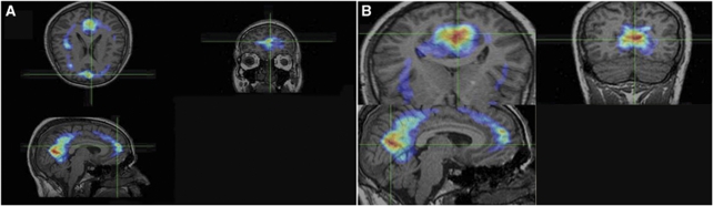
Tinnitus is often defined as the perception of sounds or noise in the absence of any external auditory stimuli. The pathophysiology of subjective idiopathic tinnitus remains unclear. The aim of this study was to investigate the functional brain activities and possible involved cerebral areas in subjective idiopathic tinnitus patients by means of single photon emission computerized tomography (SPECT) coincidence imaging, which was fused with magnetic resonance imaging (MRI). In this cross-sectional study, 56 patients (1 subject excluded) with subjective tinnitus and 8 healthy controls were enrolled. After intravenous injection of 5 mCi F18-FDG (fluorodeoxyglucose), all subjects underwent a brain SPECT coincidence scan, which was then superimposed on their MRIs. In the eight regions of interest (middle temporal, inferotemporal, medial temporal, lateral temporal, temporoparietal, frontal, frontoparietal, and parietal areas), the more pronounced values were represented in medial temporal, inferotemporal, and temporoparietal areas, which showed more important proportion of associative auditory cortices in functional attributions of tinnitus than primary auditory cortex. Brain coincidence SPECT scan, when fused on MRI is a valuable technique in the assessment of patients with tinnitus and could show the significant role of different regions of central nervous system in functional attributions of tinnitus.
Figures
References
-
- Adams PF, Hendershot GE, Marano MA. Current estimates from the National Health Interview Survey, 1996. Hyattsville, Md.: National Center for Health Statistics; 1999. - PubMed
-
- Andersson G, Lyttkens L, Hirvelä C, Furmark T, Tillfors M, Fredrikson M. Regional cerebral blood flow during tinnitus: a PET case study with lidocaine and auditory stimulation. Acta Otolaryngol. 2000;120:967–972. - PubMed
-
- Andreasen NC, Arndt S, Cizadlo T, O'Leary DS, Watkins GL, Ponto LLB, Hichwa RD. Sample size and statistical power in [15O] H2O studies of human cognition. J Cereb Blood Flow Metab. 1996;16:804–816. - PubMed
-
- Arnold W, Bartenstein P, Oesstreicher E, RÖmer W, Schwaiger M. Focal metabolic activation in the predominant left auditory cortex in patients suffering from tinnitus. A PET study with [18F] Deoxyglucose. ORL J Otorhinolaryngol Relat spec. 1996;58:195–199. - PubMed
-
- Cacace AT. Imaging tinnitus with fMRI. Assoc Res Otolaryngol Abstr. 1997;2:20.
Publication types
MeSH terms
Substances
LinkOut - more resources
Full Text Sources
Medical
