

Glinide, but not sulfonylurea, can evoke insulin exocytosis by repetitive stimulation: imaging analysis of insulin exocytosis by secretagogue-induced repetitive stimulations
- PMID: 20069052
- PMCID: PMC2801449
- DOI: 10.1155/2009/278762
Glinide, but not sulfonylurea, can evoke insulin exocytosis by repetitive stimulation: imaging analysis of insulin exocytosis by secretagogue-induced repetitive stimulations
Abstract
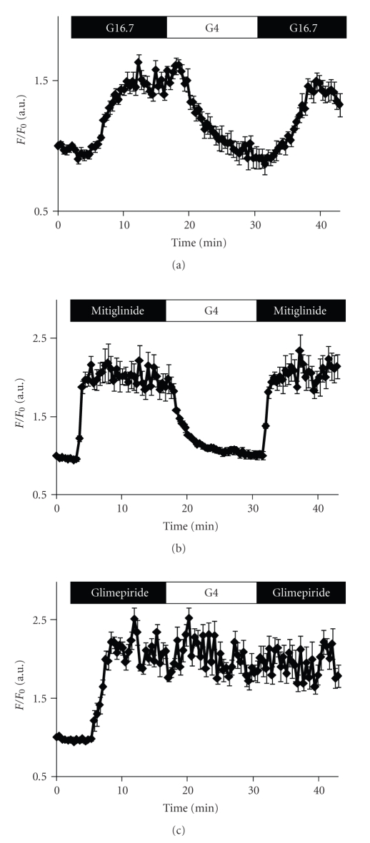
To investigate the different effects between sulfonylurea (SU) and glinide drugs in insulin secretion, pancreatic beta-cells were repeatedly stimulated with SU (glimepiride) or glinide (mitiglinide). Total internal reflection fluorescent (TIRF) microscopy revealed that secondary stimulation with glimepiride, but not glucose and mitiglinide, failed to evoke fusions of insulin granules although primary stimulation with glucose, glimepiride, and mitiglinide induced equivalent numbers of exocytotic responses. Glimepiride, but not glucose and mitiglinide, induced abnormally sustained [Ca(2+)](i) elevations and reductions of docked insulin granules on the plasma membrane. Our data suggest that the effect of glinide on insulin secretory mechanisms is similar to that of glucose.
Figures


References
-
- Quast U, Stephan D, Bieger S, Russ U. The impact of ATP-sensitive K+ channel subtype selectivity of insulin secretagogues for the coronary vasculature and the myocardium. Diabetes. 2004;53(supplement 3):S156–S164. - PubMed
-
- Kawai J, Ohara-Imaizumi M, Nakamichi Y, et al. Insulin exocytosis in Goto-Kakizaki rat β-cells subjected to long-term glinide or sulfonylurea treatment. Biochemical Journal. 2008;412(1):93–101. - PubMed
-
- Ohnota H, Koizumi T, Tsutsumi N, Kobayashi M, Inoue S, Sato F. Novel rapid- and short-acting hypoglycemic agent, a calcium(2s)-2-benzyl-3-(cis-hexahydro-2-isoindolinylcarbonyl) propionate (KAD-1229) that acts on the sulfonylurea receptor: comparison of effects between KAD-1229 and gliclazide. The Journal of Pharmacology and Experimental Therapeutics. 1994;269(2):489–495. - PubMed
-
- Karam JH, Sanz E, Salamon E, Nolte MS. Selective unresponsiveness of pancreatic B-cells to acute sulfonylurea stimulation during sulfonylurea therapy in NIDDM. Diabetes. 1986;35(12):1314–1320. - PubMed
-
- Rabuazzo AM, Buscema M, Vinci C, et al. Glyburide and tolbutamide induce desensitization of insulin release in rat pancreatic islets by different mechanisms. Endocrinology. 1992;131(4):1815–1820. - PubMed
Publication types
MeSH terms
Substances
LinkOut - more resources
Full Text Sources
Medical
Miscellaneous