Lithium chloride regulates the proliferation of stem-like cells in retinoblastoma cell lines: a potential role for the canonical Wnt signaling pathway
- PMID: 20069066
- PMCID: PMC2805422
Lithium chloride regulates the proliferation of stem-like cells in retinoblastoma cell lines: a potential role for the canonical Wnt signaling pathway
Abstract
Purpose: Cancer stem cells are found in many tumor types and are believed to lead to regrowth of tumor mass due to their chemoresistance and self-renewal capacity. We previously demonstrated small subpopulations of cells in retinoblastoma tissue and cell lines that display cancer stem cell-like activities, including expression of stem cell markers, Hoechst dye exclusion, slow cycling, and self-renewal ability. Identifying factors regulating stem cell proliferation will be important for selectively targeting stem cells and controlling tumor growth. Wingless and Int1 (Wnt) signaling is an essential cellular communication pathway that regulates proliferation and differentiation of non-neoplastic stem/progenitor cells in the retina and other tissues, but its role in cancer stem cells in the retinal tumor retinoblastoma is unknown. In this study, we investigated whether the Wnt pathway activator lithium chloride (LiCl) regulates proliferation of retinoblastoma cancer stem-like cells.
Methods: The number of stem-like cells in Weri and Y79 retinoblastoma cell line cultures was measured by 5-bromo-2-deoxyuridine (BrdU) pulse-chase, immunohistochemistry, and quantitative polymerase chain reaction (PCR) for stem cell marker genes. The cell lines were sorted into stem-like and non-stem-like populations by fluorescence-activated cell sorting (FACS), using an antibody against the stem cell marker ATP-binding cassette, subfamily G, member 2 (ABCG2). Activated Wnt signaling was measured in the sorted cells by western blotting and immunolocalization of the central mediator beta-catenin.
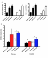
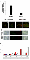
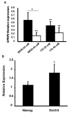
Results: LiCl increased the number of stem-like cells, measured by BrdU retention and elevated expression of the stem cell marker genes Nanog, octamer transcription factor 3 and 4 (Oct3/4), Musashi 1 (Msi1), and ABCG2. Sorted ABCG2-positive stem-like cells had higher levels of beta-catenin than ABCG2-negative non-stem cells, suggesting elevated canonical Wnt signaling. Furthermore, stem cell marker gene expression increased after small interfering RNA (siRNA) knock-down of the Wnt inhibitor secreted frizzled-related protein 2 (SFRP2).
Conclusions: These results indicate that the cancer stem-like cell population in retinoblastoma is regulated by canonical Wnt/beta-catenin signaling, which identifies the Wnt pathway as a potential mechanism for the control of stem cell renewal and tumor formation in retinoblastoma tumors in vivo.
Figures


Similar articles
-
Human embryonic and neuronal stem cell markers in retinoblastoma.Mol Vis. 2007 Jun 8;13:823-32. Mol Vis. 2007. PMID: 17615543 Free PMC article.
-
Cancer stem cell characteristics in retinoblastoma.Mol Vis. 2005 Sep 12;11:729-37. Mol Vis. 2005. PMID: 16179903
-
The Wnt/β-catenin pathway regulates self-renewal of cancer stem-like cells in human gastric cancer.Mol Med Rep. 2012 May;5(5):1191-6. doi: 10.3892/mmr.2012.802. Epub 2012 Feb 21. Mol Med Rep. 2012. PMID: 22367735
-
Wnt signaling in osteosarcoma.Adv Exp Med Biol. 2014;804:33-45. doi: 10.1007/978-3-319-04843-7_2. Adv Exp Med Biol. 2014. PMID: 24924167 Review.
-
FoxM1 and Wnt/β-catenin signaling in glioma stem cells.Cancer Res. 2012 Nov 15;72(22):5658-62. doi: 10.1158/0008-5472.CAN-12-0953. Epub 2012 Nov 8. Cancer Res. 2012. PMID: 23139209 Free PMC article. Review.
Cited by
-
β-asarone attenuates the proliferation, migration and enhances apoptosis of retinoblastoma through Wnt/β-catenin signaling pathway.Int Ophthalmol. 2023 May;43(5):1687-1699. doi: 10.1007/s10792-022-02566-1. Epub 2022 Nov 13. Int Ophthalmol. 2023. PMID: 36372820
-
Long Noncoding RNA ZFPM2-AS1 Knockdown Restrains the Development of Retinoblastoma by Modulating the MicroRNA-515/HOXA1/Wnt/β-Catenin Axis.Invest Ophthalmol Vis Sci. 2020 Jun 3;61(6):41. doi: 10.1167/iovs.61.6.41. Invest Ophthalmol Vis Sci. 2020. PMID: 32561925 Free PMC article.
-
Lithium chloride has a biphasic effect on prostate cancer stem cells and a proportional effect on midkine levels.Oncol Lett. 2016 Oct;12(4):2948-2955. doi: 10.3892/ol.2016.4946. Epub 2016 Aug 3. Oncol Lett. 2016. PMID: 27703531 Free PMC article.
-
In vitro characterization of CD133lo cancer stem cells in Retinoblastoma Y79 cell line.BMC Cancer. 2017 Nov 21;17(1):779. doi: 10.1186/s12885-017-3750-2. BMC Cancer. 2017. PMID: 29162051 Free PMC article.
-
Wnt/β-Catenin Signaling Pathway in Pediatric Tumors: Implications for Diagnosis and Treatment.Children (Basel). 2024 Jun 7;11(6):700. doi: 10.3390/children11060700. Children (Basel). 2024. PMID: 38929279 Free PMC article. Review.
References
-
- Vogelstein B, Fearon ER, Hamilton SR, Feinberg AP. Use of restriction fragment length polymorphisms to determine the clonal origin of human tumors. Science. 1985;227:642–5. - PubMed
-
- Heppner GH. Tumor heterogeneity. Cancer Res. 1984;44:2259–65. - PubMed
-
- Fidler IJ, Hart IR. Biological diversity in metastatic neoplasms: origins and implications. Science. 1982;217:998–1003. - PubMed
-
- Collins AT, Berry PA, Hyde C, Stower MJ, Maitland NJ. Prospective identification of tumorigenic prostate cancer stem cells. Cancer Res. 2005;65:10946–51. - PubMed
Publication types
MeSH terms
Substances
Grants and funding
LinkOut - more resources
Full Text Sources
Other Literature Sources
Research Materials
