

Review
Concurrent primary cardiac tumors of differing histology and origin: case report with literature review
Affiliations
- PMID: 20069087
- PMCID: PMC2801934
Item in Clipboard
Review
Concurrent primary cardiac tumors of differing histology and origin: case report with literature review
Tex Heart Inst J.
2009.
Abstract
Primary cardiac tumors are rare and are diverse in histology and anatomic origin. Approximately 75% are benign, and nearly 50% of these are myxomas. Herein, we report concurrent myxoma and papillary fibroelastoma, which tumors were found attached to the left atrial septum and aortic valve, respectively. Concurrent primary cardiac tumors of differing histology and origin are rare, and, to our knowledge, this is one of the few such cases reported in the medical literature.
Keywords: Echocardiography; fibroma; heart neoplasms/primary/diagnosis/surgery; myxoma; neoplasms, multiple primary/diagnosis/surgery.
Figures

Fig. 1. Cardiac angiography (during levophase) shows the left atrium with a filling defect that is consistent with left atrial myxoma.
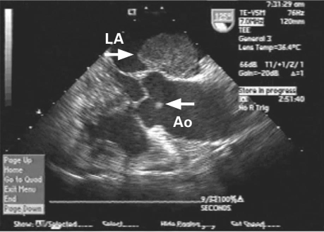
Fig. 2. Intraoperative transesophageal echocardiography (long-axis view) shows myxoma (arrow) in the left atrium (LA) and papillary fibroelastoma (arrow) attached to the noncoronary cusp of the aortic valve (Ao).

Fig. 3. Intraoperative transesophageal echocardiography (short-axis view) shows left atrial (LA) myxoma (arrow) in addition to papillary fibroelastoma (arrow) on the aortic valve (Ao).

Fig. 4. Hematoxylin and eosin stain (orig. ×200) shows a myxoid matrix dominating a hypocellular tumor. The tumor cells are in spindle, stellate, and epithelioid shapes. They are mixed with scattered lymphocytes and red blood cells.

Fig. 5. Hematoxylin and eosin stain (orig. ×40) shows papillary fibroelastoma with branching papillae, composed of central avascular collagen and variable elastic tissue, surrounded by acid mucopolysaccharide and endothelial cells.
References
-
- Straus R, Merliss S. Primary tumor of the heart. Arch Pathol 1945;39:74–8.
-
- Heath D. Pathology of cardiac tumors. Am J Cardiol 1968;21 (3):315–27. - PubMed
-
- Majano-Lainez RA. Cardiac tumors: a current clinical and pathological perspective. Crit Rev Oncog 1997;8(4):293–303. - PubMed
-
- MacGowan SW, Sidhu P, Aherne T, Luke D, Wood AE, Neligan MC, McGovern E. Atrial myxoma: national incidence, diagnosis and surgical management. Ir J Med Sci 1993;162 (6):223–6. - PubMed
Publication types
MeSH terms
LinkOut - more resources
Full Text Sources