

Isolated right superior vena cava drainage into the left atrium diagnosed noninvasively in the peripartum period
- PMID: 20069093
- PMCID: PMC2801949
Isolated right superior vena cava drainage into the left atrium diagnosed noninvasively in the peripartum period
Abstract
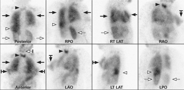
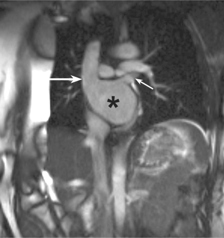
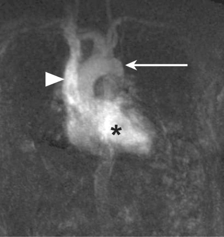
Isolated right superior vena cava drainage into the left atrium is an extremely rare cardiac anomaly, especially in the absence of other cardiac abnormalities. Only 28 of 5,127 reported consecutive congenital cardiac cases involved superior vena cava drainage into the left atrium, and all were associated with other cardiac anomalies. Of 19 reported cases of right superior vena cava drainage into the left atrium, most patients have been children who were experiencing mild hypoxemia and cyanosis. Herein, we describe the case of a 34-year-old woman who presented with asymptomatic hypoxemia in the peripartum period. She was diagnosed to have isolated drainage of the right superior vena cava into the left atrium. To the best of our knowledge, this is the 1st reported instance of such diagnosis by use of noninvasive imaging only, without cardiac catheterization. We also review the medical literature that pertains to our patient's anomaly.
Keywords: Anoxia/etiology; blood circulation; heart atria/abnormalities; heart defects, congenital/complications/diagnosis/pathology; magnetic resonance imaging; oxygen/blood; peripartum period; technetium/diagnostic use; vena cava, superior/abnormalities/radiography.
Figures
Comment in
-
Right superior caval vein draining into the left atrium.Tex Heart Inst J. 2010;37(2):255. Tex Heart Inst J. 2010. PMID: 20401313 Free PMC article. No abstract available.
References
-
- de Leval MR, Ritter DG, McGoon DC, Danielson GK. Anomalous systemic venous connection. Surgical considerations. Mayo Clin Proc 1975;50(10):599–610. - PubMed
-
- Buirski G, Jordan SC, Joffe HS, Wilde P. Superior vena caval abnormalities: their occurrence rate, associated cardiac abnormalities and angiographic classification in a paediatric population with congenital heart disease. Clin Radiol 1986; 37(2):131–8. - PubMed
-
- Park HM, Smith ET, Silberstein EB. Isolated right superior vena cava draining into left atrium diagnosed by radionuclide angiocardiography. J Nucl Med 1973;14(4):240–2. - PubMed
-
- Ezekowitz MD, Alderson PO, Bulkley BH, Dwyer PN, Watkins L, Lappe DL, et al. Isolated drainage of the superior vena cava into the left atrium in a 52-year-old man: a rare congenital malformation in the adult presenting with cyanosis, polycythemia, and an unsuccessful lung scan. Circulation 1978; 58(4):751–6. - PubMed
-
- Park HM, Summerer MH, Preuss K, Armstrong WF, Mahomed Y, Hamilton DJ. Anomalous drainage of the right superior vena cava into the left atrium. J Am Coll Cardiol 1983; 2(2):358–62. - PubMed
Publication types
MeSH terms
LinkOut - more resources
Full Text Sources
Medical