

Three-dimensional cell culture microarray for high-throughput studies of stem cell fate
- PMID: 20069558
- PMCID: PMC5333998
- DOI: 10.1002/bit.22661
Three-dimensional cell culture microarray for high-throughput studies of stem cell fate
Abstract
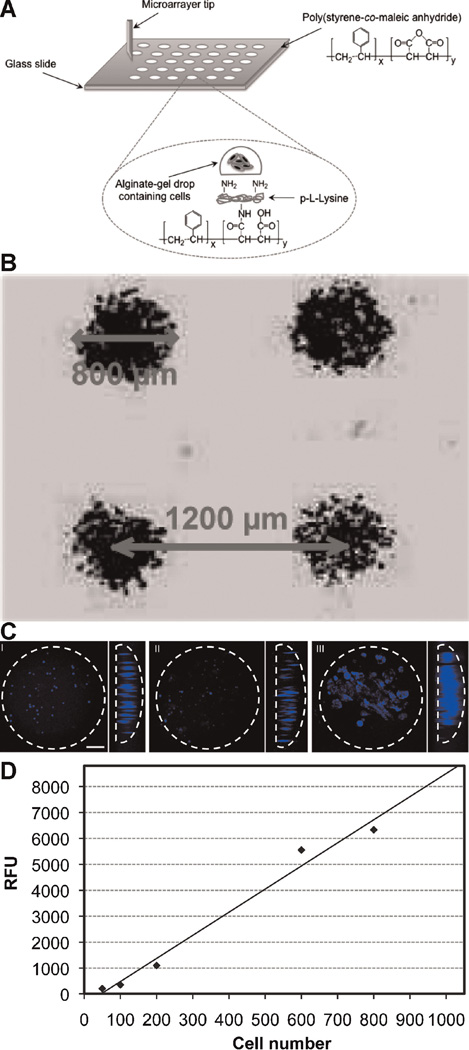
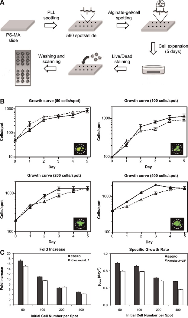
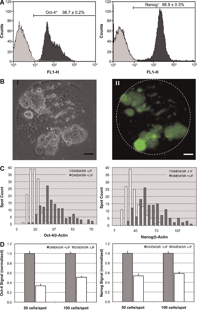
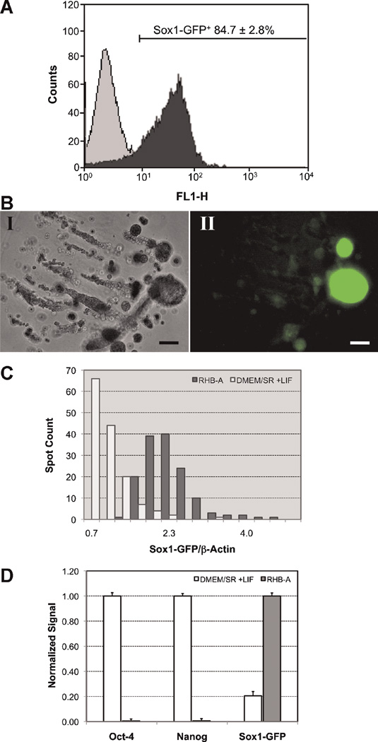
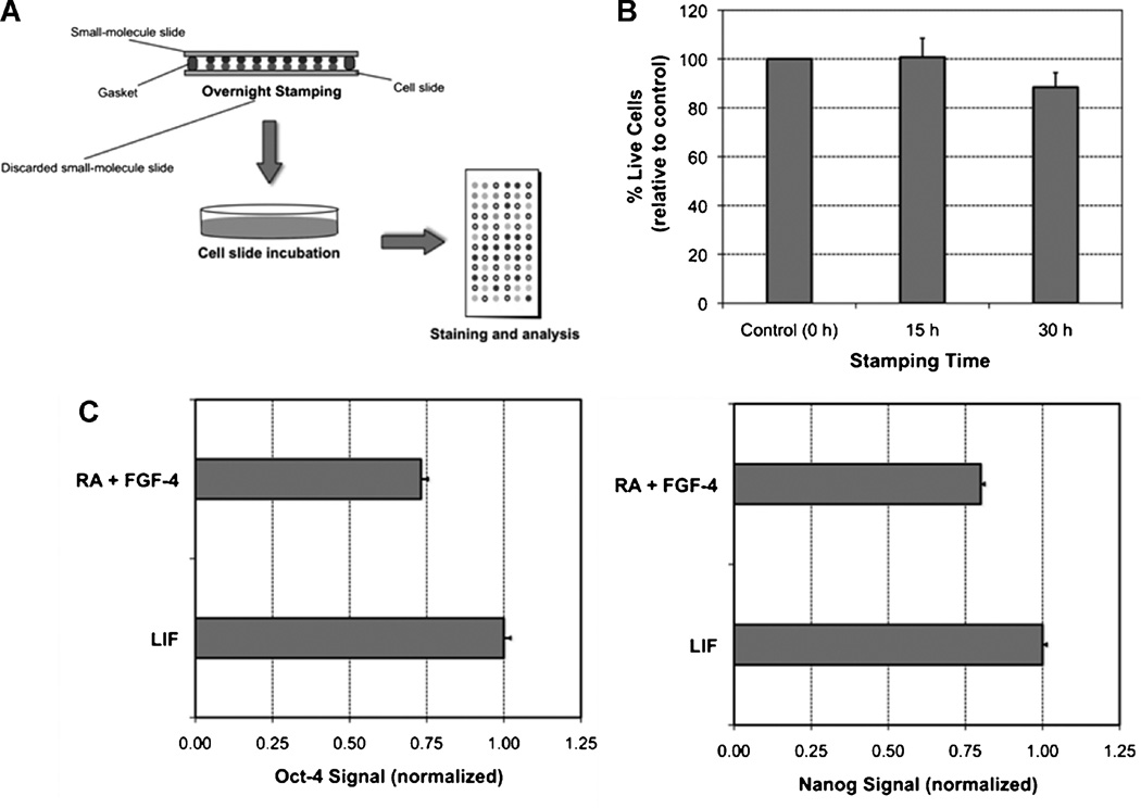
We have developed a novel three-dimensional (3D) cellular microarray platform to enable the rapid and efficient tracking of stem cell fate and quantification of specific stem cell markers. This platform consists of a miniaturized 3D cell culture array on a functionalized glass slide for spatially addressable high-throughput screening. A microarray spotter was used to deposit cells onto a modified glass surface to yield an array consisting of cells encapsulated in alginate gel spots with volumes as low as 60 nL. A method based on an immunofluorescence technique scaled down to function on a cellular microarray was also used to quantify specific cell marker protein levels in situ. Our results revealed that this platform is suitable for studying the expansion of mouse embryonic stem (ES) cells as they retain their pluripotent and undifferentiated state. We also examined neural commitment of mouse ES cells on the microarray and observed the generation of neuroectodermal precursor cells characterized by expression of the neural marker Sox-1, whose levels were also measured in situ using a GFP reporter system. In addition, the high-throughput capacity of the platform was tested using a dual-slide system that allowed rapid screening of the effects of tretinoin and fibroblast growth factor-4 (FGF-4) on the pluripotency of mouse ES cells. This high-throughput platform is a powerful new tool for investigating cellular mechanisms involved in stem cell expansion and differentiation and provides the basis for rapid identification of signals and conditions that can be used to direct cellular responses.
Figures
Similar articles
-
Three dimensional cellular microarray platform for human neural stem cell differentiation and toxicology.Stem Cell Res. 2014 Jul;13(1):36-47. doi: 10.1016/j.scr.2014.04.004. Epub 2014 Apr 18. Stem Cell Res. 2014. PMID: 24816401 Free PMC article.
-
3D-cultured neural stem cell microarrays on a micropillar chip for high-throughput developmental neurotoxicology.Exp Cell Res. 2018 Sep 15;370(2):680-691. doi: 10.1016/j.yexcr.2018.07.034. Epub 2018 Jul 24. Exp Cell Res. 2018. PMID: 30048616
-
An extracellular matrix microarray for probing cellular differentiation.Nat Methods. 2005 Feb;2(2):119-25. doi: 10.1038/nmeth736. Epub 2005 Jan 21. Nat Methods. 2005. PMID: 15782209
-
A review of gene expression profiling of human embryonic stem cell lines and their differentiated progeny.Curr Stem Cell Res Ther. 2009 May;4(2):98-106. doi: 10.2174/157488809788167409. Curr Stem Cell Res Ther. 2009. PMID: 19442194 Review.
-
Cell Microarray: An Approach to Evaluate Drug-Induced Alterations in Protein Expression.In: Sergi CM, editor. Advancements in Cancer Research [Internet]. Brisbane (AU): Exon Publications; 2023 Aug 17. Chapter 9. In: Sergi CM, editor. Advancements in Cancer Research [Internet]. Brisbane (AU): Exon Publications; 2023 Aug 17. Chapter 9. PMID: 37756425 Free Books & Documents. Review.
Cited by
-
Microfabricated biomaterials for engineering 3D tissues.Adv Mater. 2012 Apr 10;24(14):1782-804. doi: 10.1002/adma.201104631. Epub 2012 Mar 13. Adv Mater. 2012. PMID: 22410857 Free PMC article. Review.
-
Development of a high-throughput three-dimensional invasion assay for anti-cancer drug discovery.PLoS One. 2013 Dec 11;8(12):e82811. doi: 10.1371/journal.pone.0082811. eCollection 2013. PLoS One. 2013. PMID: 24349367 Free PMC article.
-
Multifactorial optimization of endothelial cell growth using modular synthetic extracellular matrices.Integr Biol (Camb). 2011 Mar;3(3):185-96. doi: 10.1039/c0ib00112k. Epub 2011 Jan 19. Integr Biol (Camb). 2011. PMID: 21249249 Free PMC article.
-
Finding the winning combination. Combinatorial screening of three dimensional niches to guide stem cell osteogenesis.Organogenesis. 2014;10(3):299-302. doi: 10.4161/org.29646. Epub 2014 Oct 31. Organogenesis. 2014. PMID: 25482315 Free PMC article.
-
Spot identification and quality control in cell-based microarrays.ACS Comb Sci. 2012 Aug 13;14(8):471-7. doi: 10.1021/co300039w. Epub 2012 Aug 1. ACS Comb Sci. 2012. PMID: 22850537 Free PMC article.
References
-
- Anderson DG, Levenberg S, Langer R. Nanoliter-scale synthesis of arrayed biomaterials and application to human embryonic stem cells. Nat Biotechnol. 2004;22:863–866. - PubMed
-
- Anderson DG, Putnam D, Lavik EB, Mahmood TA, Langer R. Biomaterial microarrays: Rapid, microscale screening of polymer-cell interaction. Biomaterials. 2005;26:4892–4897. - PubMed
-
- Castel D, Pitaval A, Debily MA, Gidrol X. Cell micrroarrays in drug discovery. Drug Discov Today. 2006;11(13/14):616–622. - PubMed
-
- Chambers I, Colby D, Robertson M, Nichols J, Lee S, Tweedie S, Smith A. Functional expression cloning of Nanog, a pluripotency sustaining factor in embryonic stem cells. Cell. 2003;113:643–655. - PubMed
Publication types
MeSH terms
Substances
Grants and funding
LinkOut - more resources
Full Text Sources
Other Literature Sources
Miscellaneous
