

Neurodevelopmental outcome of fetuses referred for ventriculomegaly
- PMID: 20069560
- PMCID: PMC2892836
- DOI: 10.1002/uog.7554
Neurodevelopmental outcome of fetuses referred for ventriculomegaly
Abstract
Objective: To characterize the delivery and postnatal neurodevelopmental outcomes of fetuses referred for ventriculomegaly (VM).
Methods: Under an internal review board-approved protocol, pregnant women were referred for magnetic resonance imaging (MRI) after sonographic diagnosis of VM and classified into one of four diagnostic groups: Group 1, normal central nervous system (CNS); Group 2, isolated mild VM (10-12 mm); Group 3, isolated VM > 12 mm; and Group 4, other CNS findings. Pregnancy outcome was obtained. Follow-up visits were offered with assessment of neurodevelopmental, adaptive and neurological functioning at 6 months and 1 year and/or 2 years of age. Atrial diameter and VM group differences in developmental outcomes were evaluated using repeated measures logistic regression and Fishers exact test, respectively.
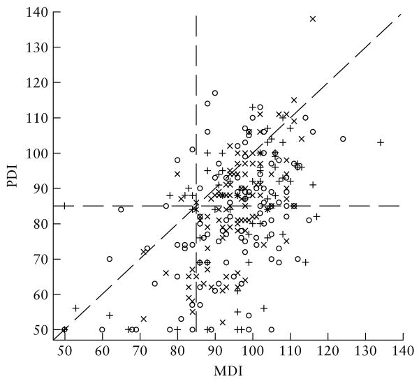
Results: Of 314 fetuses, 253 (81%) were liveborn and survived the neonatal period. Fetuses in Groups 4 and 3 were less likely to progress to live delivery and to survive the neonatal period (60% and 84%, respectively) than were those in Groups 2 or 1 (93% and 100%, respectively, P < 0.001). Of the 143 fetuses followed postnatally, between 41% and 61% had a Bayley Scales of Infant Development (BSID-II) psychomotor developmental index score in the delayed range (< 85) at the follow-up visits, whereas the BSID-II mental developmental index and Vineland Adaptive Behavior composite scores were generally in line with normative expectations. Among those that were liveborn, neither VM group nor prenatal atrial diameter was related to postnatal developmental outcome.
Conclusions: Diagnostic category and degree of fetal VM based on ultrasound and MRI measurements are associated with the incidence of live births and thus abnormal outcome. Among those undergoing formal postnatal testing, VM grade is not associated with postnatal developmental outcome, but motor functioning is more delayed than is cognitive or adaptive functioning.
Copyright 2009 ISUOG. Published by John Wiley & Sons, Ltd.
Figures
Similar articles
-
Neurological Outcome in Fetuses with Mild and Moderate Ventriculomegaly.Rev Bras Ginecol Obstet. 2016 Sep;38(9):436-442. doi: 10.1055/s-0036-1592315. Epub 2016 Sep 9. Rev Bras Ginecol Obstet. 2016. PMID: 27610601 Free PMC article.
-
Prevalence, characteristics and perinatal outcome of fetal ventriculomegaly in 29,000 pregnancies followed at a single institution.Fetal Diagn Ther. 2010;27(3):142-8. doi: 10.1159/000304735. Epub 2010 Mar 26. Fetal Diagn Ther. 2010. PMID: 20339298
-
[Fetal ventriculomegaly: diagnosis using magnetic resonance imaging and its prognosis].Zhonghua Fu Chan Ke Za Zhi. 2010 Jan;45(1):22-5. Zhonghua Fu Chan Ke Za Zhi. 2010. PMID: 20367921 Chinese.
-
Mild ventriculomegaly from fetal consultation to neurodevelopmental assessment: A single center experience and review of the literature.Eur J Paediatr Neurol. 2018 Nov;22(6):919-928. doi: 10.1016/j.ejpn.2018.04.001. Epub 2018 Apr 12. Eur J Paediatr Neurol. 2018. PMID: 29709429 Review.
-
Role of magnetic resonance imaging in fetuses with mild or moderate ventriculomegaly in the era of fetal neurosonography: systematic review and meta-analysis.Ultrasound Obstet Gynecol. 2019 Aug;54(2):164-171. doi: 10.1002/uog.20197. Epub 2019 Jul 11. Ultrasound Obstet Gynecol. 2019. PMID: 30549340
Cited by
-
Congenital Aqueductal Stenosis: Findings at Fetal MRI That Accurately Predict a Postnatal Diagnosis.AJNR Am J Neuroradiol. 2018 May;39(5):942-948. doi: 10.3174/ajnr.A5590. Epub 2018 Mar 8. AJNR Am J Neuroradiol. 2018. PMID: 29519789 Free PMC article.
-
Multi-atlas multi-shape segmentation of fetal brain MRI for volumetric and morphometric analysis of ventriculomegaly.Neuroimage. 2012 Apr 15;60(3):1819-31. doi: 10.1016/j.neuroimage.2012.01.128. Epub 2012 Feb 10. Neuroimage. 2012. PMID: 22500924 Free PMC article.
-
The Current State of Clinical Trials Studying Hydrocephalus: An Analysis of ClinicalTrials.gov.Cureus. 2020 Aug 25;12(8):e10029. doi: 10.7759/cureus.10029. Cureus. 2020. PMID: 32983722 Free PMC article.
-
Cortical folding alterations in fetuses with isolated non-severe ventriculomegaly.Neuroimage Clin. 2018 Jan 28;18:103-114. doi: 10.1016/j.nicl.2018.01.006. eCollection 2018. Neuroimage Clin. 2018. PMID: 29387528 Free PMC article.
-
Early alterations in cortical and cerebellar regional brain growth in Down Syndrome: An in vivo fetal and neonatal MRI assessment.Neuroimage Clin. 2020;25:102139. doi: 10.1016/j.nicl.2019.102139. Epub 2019 Dec 23. Neuroimage Clin. 2020. PMID: 31887718 Free PMC article.
References
-
- Garel C, Luton RJ. Ventricular dilatations. Childs Nerv Syst. 2003;19:301–308. - PubMed
-
- Nicolaides KH, Berry S, Snijders RJM, Thorpe-Beeston JG, Gosden C. Fetal lateral cerebral ventriculomegaly: Associated malformations and chromosomal defects. Fetal Diagn Ther. 1990;5:5–14. - PubMed
-
- Goldstein I, Reece EA, Pilu GL. Sonographic evaluation of the normal developmental anatomy of the fetal cerebral ventricles. IV. The posterior horn. Am J Perinatol. 1990;7:79–83. - PubMed
-
- Bromley B, Frigoletto FD, Jr, Benacerraf BR. Mild fetal lateral cerebral ventriculomegaly: clinical course and outcome. Am J Obstet Gynecol. 1991;164:863–867. - PubMed
-
- Bloom SL, Bloom DD, DellaNebbia C, Martin LB, Lucas MJ, Twickler DM. The developmental outcome of children with antenatal mild isolated ventriculomegaly. Obstet Gynecol. 1997;90:93–97. - PubMed
Publication types
MeSH terms
Grants and funding
LinkOut - more resources
Full Text Sources
Other Literature Sources
Medical
