. 2010 Jun 1;12(11):1235-46.
doi: 10.1089/ars.2009.2701.

Irreversible inactivation of glutathione peroxidase 1 and reversible inactivation of peroxiredoxin II by H2O2 in red blood cells

Affiliations

Irreversible inactivation of glutathione peroxidase 1 and reversible inactivation of peroxiredoxin II by H2O2 in red blood cells

Chun-Seok Cho et al. Antioxid Redox Signal. .

Abstract

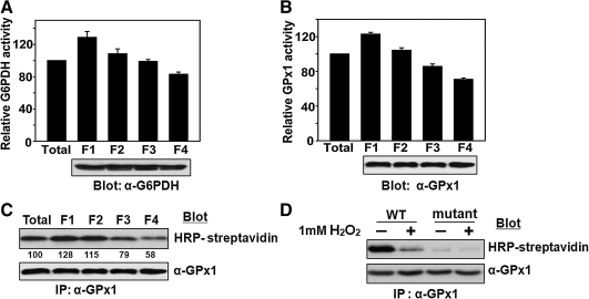
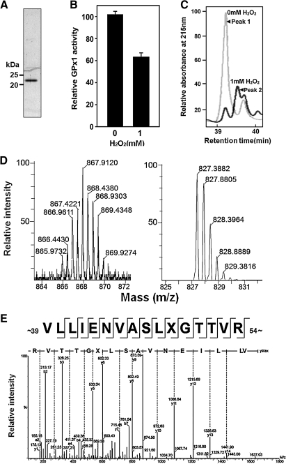
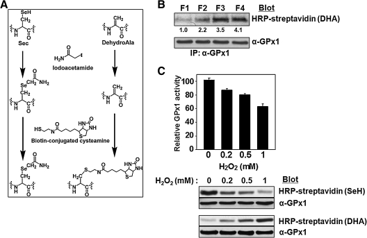
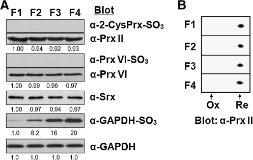
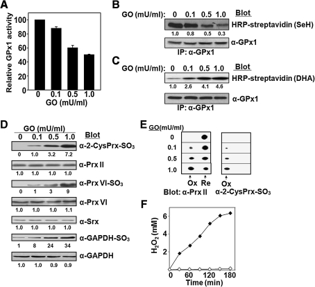
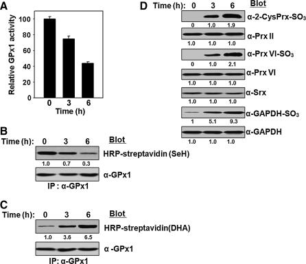
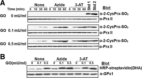
Catalase, glutathione peroxidase1 (GPx1), and peroxiredoxin (Prx) II are the principal enzymes responsible for peroxide elimination in RBC. We have now evaluated the relative roles of these enzymes by studying inactivation of GPx1 and Prx II in human RBCs. Mass spectrometry revealed that treatment of GPx1 with H(2)O(2) converts the selenocysteine residue at its active site to dehydroalanine (DHA). We developed a blot method for detection of DHA-containing proteins, with which we observed that the amount of DHA-containing GPx1 increases with increasing RBC density, which is correlated with increasing RBC age. Given that the conversion of selenocysteine to DHA is irreversible, the content of DHA-GPx1 in each RBC likely reflects total oxidative stress experienced by the cell during its lifetime. Prx II is inactivated by occasional hyperoxidation of its catalytic cysteine to cysteine sulfinic acid during catalysis. We believe that the activity of sulfiredoxin in RBCs is sufficient to counteract the hyperoxidation of Prx II that occurs in the presence of the basal level of H(2)O(2) flux resulting from hemoglobin autoxidation. If the H(2)O(2) flux is increased above the basal level, however, the sulfinic Prx II begins to accumulate. In the presence of an increased H(2)O(2) flux, inhibition of catalase accelerated the accumulation of sulfinic Prx II, indicative of the protective role of catalase.


Figures

FIG. 1.
FIG. 1.
Effect of aging on the catalytic activity and selenol content of GPx1 in RBCs. (A–C) Fresh RBCs obtained from a healthy human adult were separated on the basis of their density by centrifugation at 4,000 g for 15 min at 4°C on a discontinuous density gradient consisting of 85, 80, 76, 72, 69, and 66% Percoll (from the bottom up), as described previously (8). Among the six discrete bands obtained, the two minor bands at the top and bottom were discarded and the four middle bands (F1 to F4 for the least dense to the most dense, respectively) were used. The activity of G6PDH (A) in each fraction were measured according to the procedure described previously (29), and the activity of GPx1 (B) were measured, and their activities were normalized by the corresponding value for RBCs before fractionation. The fractions were also assayed for the selenol content of GPx1 (C); RBC lysates were subjected to alkylation with BIAM, GPx1 was immunoprecipitated (IP) from the lysates with antibodies to GPx1, and BIAM-labeled GPx1 in the precipitates was detected by blot analysis with HRP-conjugated streptavidin to measure selenol content (SeH). The band intensities are the average of three independent experiments. The presence of equal amounts of protein among assay mixtures was confirmed by immunoblot analysis with antibodies to G6PDH (A) or to GPx1 (B, C). Activity data in A and B are means ± SD of triplicates from a representative experiment. (D) Wild-type (WT) and the Sec49 → Cys mutant (mutant) GPx1(10 μg) were incubated in the absence or presence of 1 mM H2O2 for 1 h at 37°C and precipitated with trichloroacetic acid. The precipitated proteins were subjected to BIAM labeling, followed by HRP-conjugated streptavidin blot analysis as described in C.
FIG. 2.
FIG. 2.
Identification of DHA in H2O2-treated GPx1 by MS. (A) Purified human GPx1 was subjected to SDS-PAGE analysis on a 14% gel. Size markers are indicated. (B) Purified Gpx1 (30 μg) was incubated in the absence or presence of 1 mM H2O2 for 1 h at 37°C and then assayed for GPx1 activity. Data are means ± SD of triplicates from a representative experiment. (C) GPx1 incubated with (dark gray line) or without (light gray line) H2O2 as in B was subjected to tryptic digestion, and the resulting peptides were fractionated by HPLC on a C18 column. Peaks eluting between 39 and 40 min are shown. (D) Expanded LC-ESI-Q-TOF tandem MS spectra for peptides corresponding to peak 1 (left panel) or peak 2 (right panel) from C. The isotopic distribution was normalized relative to the largest peak. (E) Tandem MS spectrum obtained from fragmentation of the doubly charged ion with an m/z of 827.4 from peak 2 in D. The y ion series defined the indicated amino acid sequence. The mass difference of 69.0 Da between the y5 and y6 ions defined residue X as DHA.
FIG. 3.
FIG. 3.
Effect of aging and H2O2 treatment on the DHA content of GPx1 as revealed by blot analysis after reaction with biotin-conjugated cysteamine. (A) Chemical reactions underlying the biotinylation of GPx1 containing DHA. After alkylation of free SH and SeH groups by iodoacetamide, DHA residues are biotinylated with biotin-conjugated cysteamine. (B) Fresh RBCs were separated into four fractions (F1, F2, F3, and F4) on the basis of their density (age) as in Fig. 1, and GPx1 was immunoprecipitated from the lysate of each fraction and analyzed for DHA content by sequential reaction with iodoacetamide and biotin-conjugated cysteamine as outlined in A. The band intensities are average of three independent experiments. (C) After incubation of purified GPx1 (10 μg) at 37°C for 1 h with various amounts (0, 0.2, 0.5, or 1 mM ) of H2O2, GPx activity, selenol content(SeH), and DHA-content were measured as described above.
FIG. 4.
FIG. 4.
Effects of aging on generation of the sulfinic forms of Prx enzymes or GAPDH. (A) Fresh RBCs were separated into four fractions (F1, F2, F3, and F4) on the basis of their density (age), and lysates of each fraction were subjected to immunoblot analysis with antibodies specific for the sulfinic form of 2-Cys Prxs, Prx VI, or GAPDH. Equal loading of proteins was confirmed by immunoblot analysis with antibodies to Prx II, to Prx VI, and to GAPDH, respectively. Immunoblot analysis was also performed with antibodies to Srx. The band intensities are average of two independent experiments. (B) Lysates of the age-related fractions of RBCs were also subjected to two-dimensional PAGE on a 13-cm Immobiline DryStrips (Amersham; pH 4 to 7, linear) and probed by immunoblot analysis with antibodies to Prx II(20). The positions of hyperoxidized (Ox) and reduced (Re) forms of Prx II are indicated.
FIG. 5.
FIG. 5.
Effects on antioxidant enzymes in RBCs of extracellular H2O2 produced by GO. (A–E) Various amounts (0, 0.1, 0.5, or 1 mU) of glucose oxidase (GO) were added to 1 ml of RBCs at a 50% hematocrit in DMEM containing a high concentration (4,500 mg/l) of glucose. After incubation for 3 h with gentle shaking at 37°C, the RBCs were lysed and subjected to the following analyses: (A) Measurement of GPx1 activity and its normalization by that of RBCs incubated in the absence of GO. Data are means ± SD of triplicates from a representative experiment. (B, C) Determination of the selenol (SeH) and DHA contents, respectively, of GPx1. Equal loading of proteins was confirmed by immunoblot analysis with antibodies to GPx1. (D) Immunoblot analysis with antibodies specific for the sulfinic forms of 2-Cys Prxs, Prx VI, or GAPDH. Equal loading of proteins was confirmed by immunoblot analysis with antibodies to Prx II, to Prx VI, or to GAPDH, respectively. Immunoblot analysis was also performed with antibodies to Srx. The band intensities shown in BD are average of two independent experiments. (E) Two-dimensional PAGE followed by immunoblot analysis with antibodies to Prx II and to the sulfinic form of 2-Cys Prxs. The positions of hyperoxidized (Ox) and reduced (Re) forms of Prx II are indicated. (F) The H2O2 concentration generated by incubation of GO at 1 mU/ml in the absence (solid diamonds) or presence (open diamonds) of RBCs at a 50% hematocrit was measured on the basis of ferrous oxidation of xylenol orange (45).
FIG. 6.
FIG. 6.
Effects of N-phenylhydroxylamine on antioxidant enzymes in RBCs. N-phenylhydroxy-lamine (200 μM) was added to a 50% hematocrit of RBCs in DMEM containing a high glucose concentration (4500 mg/l). After incubation for various times (0, 3, or 6 h) with gentle shaking at 37°C, the RBCs were lysed and subjected to the following analyses: (A) Measurement of GPx1 activity and its normalization relative to that of cells incubated in the absence of N-phenylhydroxylamine (time 0). Data are means ± SD of triplicates from a representative experiment. (B, C) Determination of the selenol (SeH) and DHA contents, respectively, of GPx1. Equal loading of proteins was confirmed by immunoblot analysis with antibodies to GPx1. (D) Immunoblot analysis with antibodies specific for the sulfinic forms of 2-Cys Prxs, Prx VI, or GAPDH. Equal loading of proteins was confirmed by immunoblot analysis with antibodies to Prx II, to Prx VI, and to GAPDH, respectively. Immunoblot analysis was also performed with antibodies to Srx. The band intensities shown in BD are the average of two independent experiments.
FIG. 7.
FIG. 7.
Effect of catalase inhibition on Prx II hyperoxidation and GPx1-DHA formation. RBCs at a 50% hematocrit in DMEM containing high glucose (4500 mg/l) were incubated at 37°C first for 10 min in the absence or presence of 10 mM sodium azide or 50 mM 3-AT and then for the indicated times (0, 10, 30, and 60 min) in the additional presence of various concentrations (0, 0.1, or 0.5 mU/ml) of GO. (A) The cells were then lysed and subjected to immunoblot analysis with antibodies specific for the sulfinic form of 2-Cys Prxs. Equal loading of proteins was confirmed by immunoblot analysis with antibodies to Prx II. Lysates of RBCs that had been treated with GO at 0.1 mU/ml (Std 1) or 0.5 mU/ml (Std 2) for 3 h (Fig. 5E) were included in the immunoblot analysis as standards; in these standard samples, ∼10 and ∼25% of Prx II was hyperoxidized as judged on the basis of 2D-PAGE analysis. (B) RBCs that had been incubated for 60 min with various concentrations (0, 0.1, or 0.5 mU/ml) of GO in the absence or presence of 10 mM sodium azide or 50 mM 3-AT as in A were lysed, and the lysates were then subjected to the determination of DHA contents. Equal loading of proteins was confirmed by immunoblot analysis with antibodies to GPx1.

Similar articles

Cited by

References

    1. Andersen HR. Nielsen JB. Nielsen F. Grandjean P. Antioxidative enzyme activities in human erythrocytes. Clin Chem. 1997;43:562–568. - PubMed
    1. Awasthi YC. Beutler E. Srivastava SK. Purification and properties of human erythrocyte glutathione peroxidase. J Biol Chem. 1975;250:5144–5149. - PubMed
    1. Bartosz G. Aging of the erythrocyte. VII. On the possible causes of inactivation of red cell enzymes. Mech Ageing Dev. 1980;13:379–385. - PubMed
    1. Beutler E. The hemolytic effect of primaquine and related compounds: A review. Blood. 1959;14:103–139. - PubMed
    1. Biteau B. Labarre J. Toledano MB. ATP-dependent reduction of cysteine-sulphinic acid by S. cerevisiae sulphiredoxin. Nature. 2003;425:980–984. - PubMed

Publication types

MeSH terms

LinkOut - more resources