

Integrated pathways for neutrophil recruitment and inflammation in leprosy
- PMID: 20070238
- PMCID: PMC2811231
- DOI: 10.1086/650318
Integrated pathways for neutrophil recruitment and inflammation in leprosy
Abstract
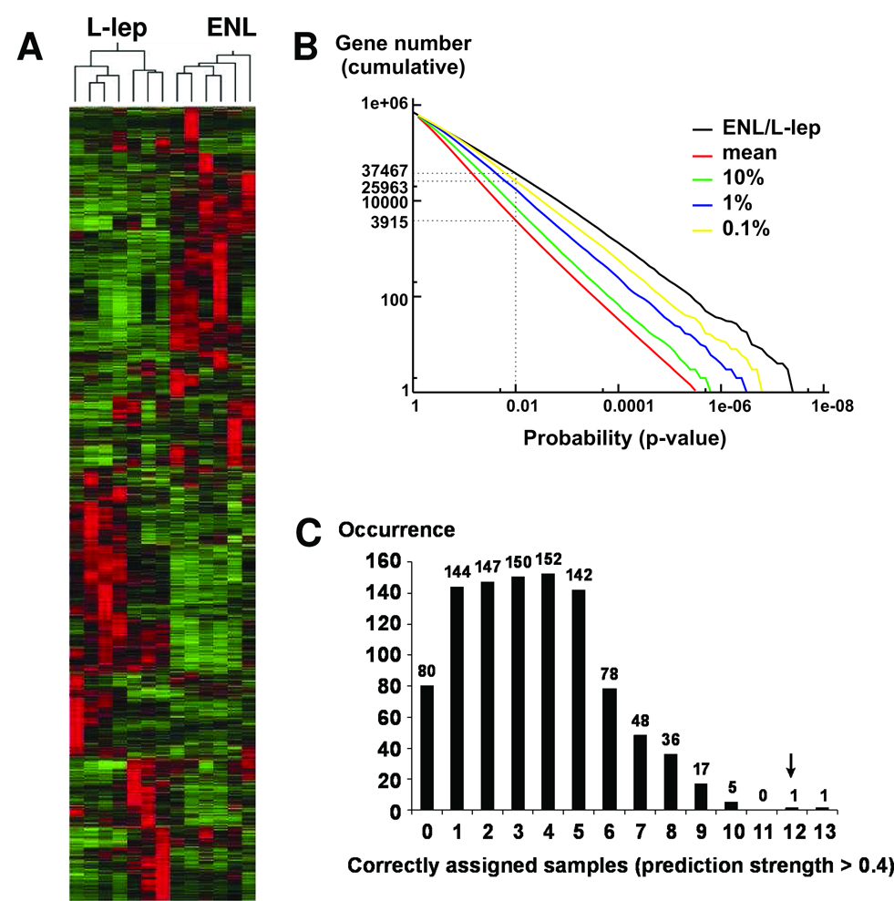
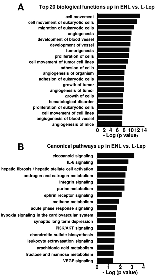
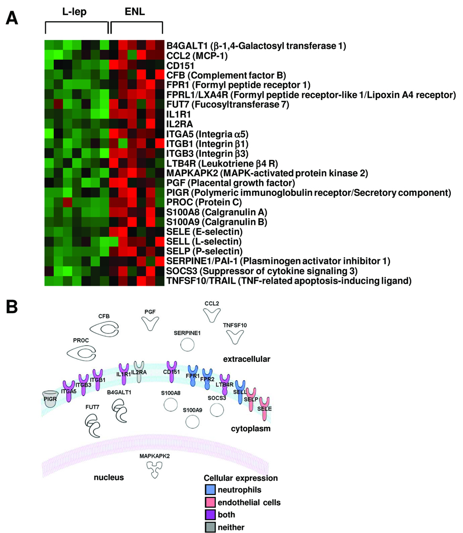
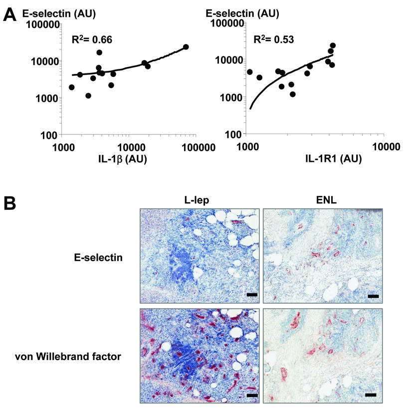
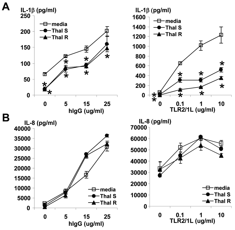
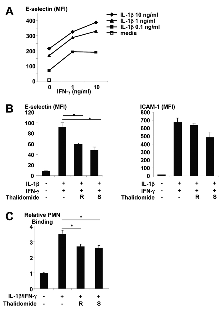
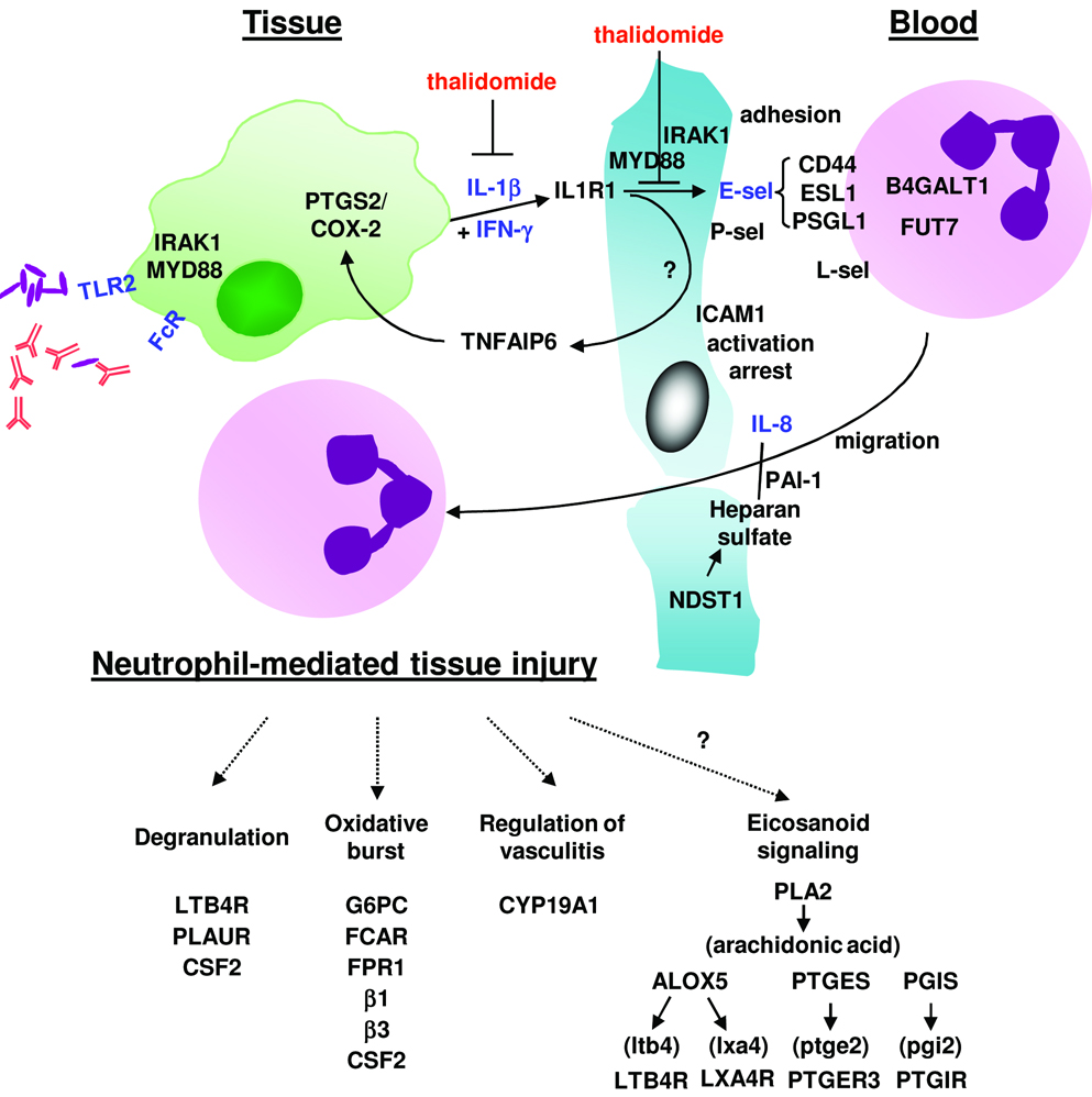
Neutrophil recruitment is pivotal to the host defense against microbial infection, but it also contributes to the immunopathology of disease. We investigated the mechanism of neutrophil recruitment in human infectious disease by means of bioinformatic pathways analysis of the gene expression profiles in the skin lesions of leprosy. In erythema nodosum leprosum (ENL), which occurs in patients with lepromatous leprosy and is characterized by neutrophil infiltration in lesions, the most overrepresented biological functional group was cell movement, including E-selectin, which was coordinately regulated with interleukin 1beta (IL-1beta). In vitro activation of Toll-like receptor 2 (TLR2), up-regulated in ENL lesions, triggered induction of IL-1beta, which together with interferon gamma induced E-selectin expression on and neutrophil adhesion to endothelial cells. Thalidomide, an effective treatment for ENL, inhibited this neutrophil recruitment pathway. The gene expression profile of ENL lesions comprised an integrated pathway of TLR2 and Fc receptor activation, neutrophil migration, and inflammation, providing insight into mechanisms of neutrophil recruitment in human infectious disease.
Conflict of interest statement
Potential conflicts of interest: none.
Figures
Similar articles
-
Whole blood transcriptomics reveals the enrichment of neutrophil activation pathways during erythema nodosum leprosum reaction.Front Immunol. 2024 Apr 23;15:1366125. doi: 10.3389/fimmu.2024.1366125. eCollection 2024. Front Immunol. 2024. PMID: 38715615 Free PMC article.
-
Differential Expression of IFN-γ, IL-10, TLR1, and TLR2 and Their Potential Effects on Downgrading Leprosy Reaction and Erythema Nodosum Leprosum.J Immunol Res. 2019 Nov 7;2019:3405103. doi: 10.1155/2019/3405103. eCollection 2019. J Immunol Res. 2019. PMID: 31781675 Free PMC article.
-
Erythema Nodosum Leprosum Neutrophil Subset Expressing IL-10R1 Transmigrates into Skin Lesions and Responds to IL-10.Immunohorizons. 2020 Feb 7;4(2):47-56. doi: 10.4049/immunohorizons.1900088. Immunohorizons. 2020. PMID: 32034084
-
Neutrophils in Leprosy.Front Immunol. 2019 Mar 19;10:495. doi: 10.3389/fimmu.2019.00495. eCollection 2019. Front Immunol. 2019. PMID: 30949168 Free PMC article. Review.
-
Thalidomide in the treatment of leprosy.Microbes Infect. 2002 Sep;4(11):1193-202. doi: 10.1016/s1286-4579(02)01645-3. Microbes Infect. 2002. PMID: 12361920 Review.
Cited by
-
Host-Related Laboratory Parameters for Leprosy Reactions.Front Med (Lausanne). 2021 Oct 22;8:694376. doi: 10.3389/fmed.2021.694376. eCollection 2021. Front Med (Lausanne). 2021. PMID: 34746168 Free PMC article. Review.
-
Murine experimental leprosy: Evaluation of immune response by analysis of peritoneal lavage cells and footpad histopathology.Int J Exp Pathol. 2019 Jun;100(3):161-174. doi: 10.1111/iep.12319. Epub 2019 May 24. Int J Exp Pathol. 2019. PMID: 31124597 Free PMC article.
-
Whole blood transcriptomics reveals the enrichment of neutrophil activation pathways during erythema nodosum leprosum reaction.Front Immunol. 2024 Apr 23;15:1366125. doi: 10.3389/fimmu.2024.1366125. eCollection 2024. Front Immunol. 2024. PMID: 38715615 Free PMC article.
-
What is New in the Pathogenesis and Management of Erythema Nodosum Leprosum.Indian Dermatol Online J. 2020 Jul 13;11(4):482-492. doi: 10.4103/idoj.IDOJ_561_19. eCollection 2020 Jul-Aug. Indian Dermatol Online J. 2020. PMID: 32832433 Free PMC article. Review.
-
Neutrophil-derived IL-1β is sufficient for abscess formation in immunity against Staphylococcus aureus in mice.PLoS Pathog. 2012;8(11):e1003047. doi: 10.1371/journal.ppat.1003047. Epub 2012 Nov 29. PLoS Pathog. 2012. PMID: 23209417 Free PMC article.
References
-
- Miller LS, O'Connell RM, Gutierrez MA, et al. MyD88 mediates neutrophil recruitment initiated by IL-1R but not TLR2 activation in immunity against Staphylococcus aureus. Immunity. 2006 Jan;24(1):79–91. - PubMed
-
- Xu Q, Seemanapalli SV, Reif KE, Brown CR, Liang FT. Increasing the recruitment of neutrophils to the site of infection dramatically attenuates Borrelia burgdorferi infectivity. J Immunol. 2007 Apr 15;178(8):5109–5115. - PubMed
-
- Movat HZ. Tissue injury and inflammation induced by immune complexes: the critical role of the neutrophil leukocyte. Exp Mol Pathol. 1979 Aug;31(1):201–210. - PubMed
-
- Anthony J, Vaidya MC, Dasgupta A. Ultrastructure of skin in erythema nodosum leprosum. Cytobios. 1983;36(141):17–23. - PubMed
Publication types
MeSH terms
Substances
Grants and funding
LinkOut - more resources
Full Text Sources
Medical
Molecular Biology Databases
