Review
. 2010 Jan 15;425(3):501-12.
doi: 10.1042/BJ20090861.

Taking the plunge: integrating structural, enzymatic and computational insights into a unified model for membrane-immersed rhomboid proteolysis

Affiliations
Review

Taking the plunge: integrating structural, enzymatic and computational insights into a unified model for membrane-immersed rhomboid proteolysis

Sinisa Urban. Biochem J. .

Abstract

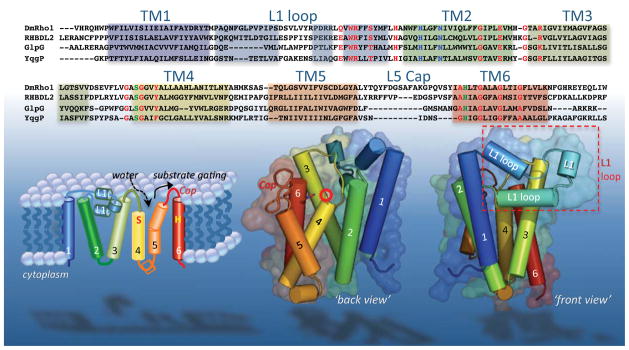
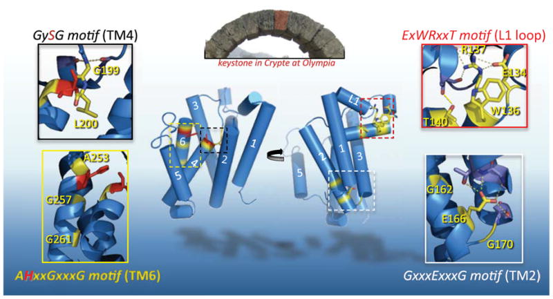
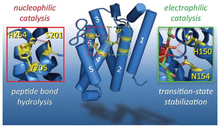
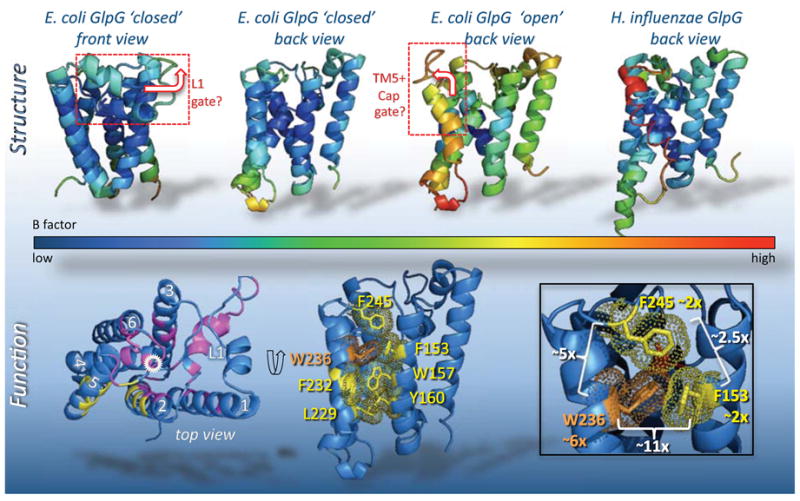
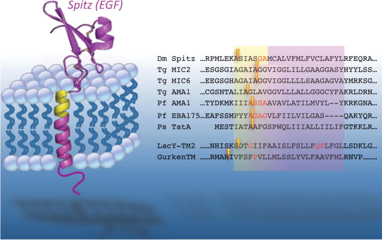
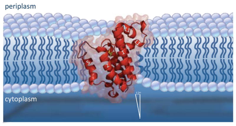
Rhomboid proteases are a fascinating class of enzymes that combine a serine protease active site within the core of an integral membrane protein. Despite having key roles in animal cell signalling and microbial pathogenesis, the membrane-immersed nature of these enzymes had long imposed obstacles to elucidating their biochemical mechanisms. But recent multidisciplinary approaches, including eight crystal structures, four computer simulations and nearly 100 engineered mutants interrogated in vivo and in vitro, are coalescing into an integrated model for one rhomboid orthologue in particular, bacterial GlpG. The protein creates a central hydrated microenvironment immersed below the membrane surface to support hydrolysis by its serine protease-like catalytic apparatus. Four conserved architectural elements in particular act as 'keystones' to stabilize this structure, and the lateral membrane-embedded L1 loop functions as a 'flotation device' to position the protease tilted in the membrane. Complex interplay between lateral substrate gating by rhomboid, substrate unwinding and local membrane thinning leads to intramembrane proteolysis of selected target proteins. Although far from complete, studies with GlpG currently offer the best prospect for achieving a thorough and sophisticated understanding of a simplified intramembrane protease.


Figures

Figure 1
Figure 1. Sequence and structural features of the rhomboid transmembrane core
Top panel lists all residues used in the E. coli GlpG structure analyses (residues 87–276), aligned with the corresponding regions of three other well-studied rhomboid enzymes (rhomboid-1 from Drosophila, human RHBDL2, and B. subtilis YqgP). As such, amino terminal domains of all enzymes, and the seventh TM and carboxy terminal domains of DmRho1, RHBDL2 and YqgP are not shown. Residues involved in nucleophilic catalysis (S201 and H254 for GlpG) are shaded in green, presumed electrophilic catalysis residues (H150 and N154 for GlpG) are shaded in blue, while conserved structural residues are shaded in red. Helical secondary structure is highlighted by common color shading among the sequence alignment (top), cylinders in the topology diagram (lower left), and crystal structure images (lower middle and right). Note the intramembrane location of the catalytic serine (circled with red star), and the protruding L1 loop (boxed lower right). By convention, the ‘back view’ centers on TM5 and TM2, while the ‘front view’ centers on TM1 and the L1 loop.
Figure 2
Figure 2. Four keystones of E. coli GlpG rhomboid protease structure
The central stone in an arch is called a keystone, and although arch stability depends on many factors, the keystone occupies a key, central stabilizing position. Pictured in the top inset is the Crypte at Olympia, Greece, with the keystone false-colored red (photo: S. Urban). The back (left) and front (right) views of GlpG are shown. By analogy, ‘keystone’ residues (highlighted in yellow, active site residues in red) of four conserved motifs are depicted in each inset. From top right and moving clockwise: the sidechain of R137 of the ExWRxxT motif reaches upwards to make 5 hydrogen bonds within the L1 loop, two to E134, and three to backbone carbonyls of L121 and A124, while T140 hydrogen bonds to the carbonyl of L123. This array of interactions together stabilizes the lateral hairpin. The E166 sidechain of the GxxxExxxG motif in TM2 makes four hydrogen bonds: three to backbone and sidechain atoms of V96 and T97 in TM1, and one to the hydroxyl of S171 in TM3, bringing the three TMs into an apex on the cytosolic side (bottom). TM2 and the L2 loop bend around G162 and G170, respectively. G199 in the GySG motif of TM4 hydrogen-bonds with the backbone carbonyls of H141 and M144 of the L1 loop, while L200 is buried into the ‘floor’ of the enzyme. AHxxGxxxG motif in TM6 mediates close approach of TM6 with TM3 (via A253), and with TM4 (via G257 and G261). Oxygen atoms are in red, nitrogen atoms are in blue, and dashed lines depict hydrogen bonds throughout.
Figure 3
Figure 3. The two catalytic elements of the GlpG rhomboid protease active site
Nucleophilic catalysis (left) that is responsible for severing the peptide bond is carried out by a serine protease-like catalytic dyad: S201 (on TM4) serves as the nucleophile while the catalytic base is H254 (on TM6). H254 hydrogen-bonding to S201 is depicted with a dashed red line. The phenol ring of Y205, emanating from TM4, stacks under the H254 imidazole ring, providing some support for the catalytic base (although a tyrosine could not salt-bridge with the imidazolium ion or alter its pKa, as might occur in some proteases). Electrophilic catalysis (right) is required to stabilize the oxyanion of the tetrahedral intermediate. The residues that form this entity are less defined, but are suggested by interaction with a lipid phosphate that protrudes into the active site of structure 2IRV. The lipid is shown in green, with oxygen atoms in red. Hydrogen bonds between the phosphate oxygen and the sidechains of H150 and N154 (emanating from TM2) are depicted by red dashed lines.
Figure 4
Figure 4. Structural and functional analyses of GlpG substrate gating
Models for substrate entry posited either lifting away of the L1 loop (boxed in top left), or displacement of TM5 and the overlying Cap (shown already in the open position). In addition to the conformational differences between structures, B factors also serve indirectly to suggest regions of conformational flexibility. The three structures are colored by B factor values (see underlying spectrum). In the first structure (2IC8, leftmost two views), the B factors are relatively low throughout, while in subsequent structures the higher B factors tend to center on TM5 and connecting loops. Bottom panels summarize effects of GlpG mutants on protease activity from a screen to identify gating residues [41]; from an analysis of ~50 mutants engineered in all TMs and outer loops, mutants that increased protease activity only localized in the TM5, TM2 and Cap interface (in yellow). Locations of targeted mutants that did not increase activity are shown in magenta (catalytic serine is circled in white). In the middle lower panel, the interface that prohibits substrate access is shown in the ‘closed’ form, with protruding sidechains and their electron density in yellow or orange (for W236). The lower right panel magnifies the interface formed by W236 (TM5), F245 (Cap) and F153 (TM2). The greatest stimulatory effect of any single mutant within GlpG occurs when W236 (shown in orange) is mutated to alanine, resulting in a ~6-fold (~6x) increase in enzyme activity, compared to an approximate doubling (~2x) when either F153 or F245 are mutated to alanine. White brackets summarize the increase in protease activity relative to wildtype of all pairwise double mutants [42]: note that only the F153A+W236A mutant combination is synergistic, resulting in a ~11-fold increase in protease activity.
Figure 5
Figure 5. Substrate requirements for rhomboid processing
The first identified rhomboid substrate, the Drosophila EGF ligand Spitz, is depicted in the membrane. Note that structural information is available for only the EGF domain. The ‘substrate motif’ in Spitz that is necessary and sufficient for rhomboid cleavage is highlighted in yellow. The right panel shows all of the cleavage sites, depicted with lightning bolts, currently known for different rhomboid protease substrates (excluding the mitochondrial rhomboid substrates). Approximate transmembrane and substrate regions are depicted, while residues experimentally determined to act as helix-breaking are in red font. The cleavage sites for the top seven substrates were determined with natural proteins, while the bottom two are artificial substrates. Note that in natural substrates the cleavage site always follows an alanine residue. Organism designations are: Dm Drosophila, Tg Toxoplasma gondii, Pf Plasmodium falciparum, Ps Providencia stuartii. The artificial substrates are chimeric proteins harbouring the transmembrane segments from the E. coli LacY permease and Drosophila Gurken.
Figure 6
Figure 6. GlpG in a membrane: protease tilt and membrane thinning
A stylized diagram of GlpG tilt and membrane thinning as suggested by molecular dynamics simulations. Interactions with membrane lipids, including through the L1 loop acting as a ‘flotation device’, serve to stabilize GlpG in a position tilted ~12 degrees from the membrane normal. This results in slightly deeper submersion of the catalytic serine (in yellow) and TM5. The hydrophobic mismatch between GlpG and membrane also causes deformation of the membrane in the immediate vicinity of GlpG, resulting in a ~4A thinner lipid annulus. The most extreme thinning occurs on the cytosolic side under the L1 loop. Enzyme tilt and membrane thinning may play roles in how substrates approach and are handled by rhomboid proteases.

References

    1. Lopez-Otin C, Overall CM. Protease degradomics: a new challenge for proteomics. Nat Rev Mol Cell Biol. 2002;3:509–519. - PubMed
    1. Lopez-Otin C, Bond JS. Proteases: multifunctional enzymes in life and disease. J Biol Chem. 2008;283:30433–30437. - PMC - PubMed
    1. Polgar L. The catalytic triad of serine peptidases. Cell Mol Life Sci. 2005;62:2161–2172. - PMC - PubMed
    1. Blow DM, Birktoft JJ, Hartley BS. Role of a buried acid group in the mechanism of action of chymotrypsin. Nature. 1969;221:337–340. - PubMed
    1. Blow D. So do we understand how enzymes work? Structure. 2000;8:R77–81. - PubMed

Publication types

LinkOut - more resources