. 2010 Mar;216(3):407-16.
doi: 10.1111/j.1469-7580.2009.01190.x. Epub 2010 Jan 7.

Distribution and expression of CD200 in the rat respiratory system under normal and endotoxin-induced pathological conditions

Affiliations

Distribution and expression of CD200 in the rat respiratory system under normal and endotoxin-induced pathological conditions

Ya-Fen Jiang-Shieh et al. J Anat. 2010 Mar.

Abstract

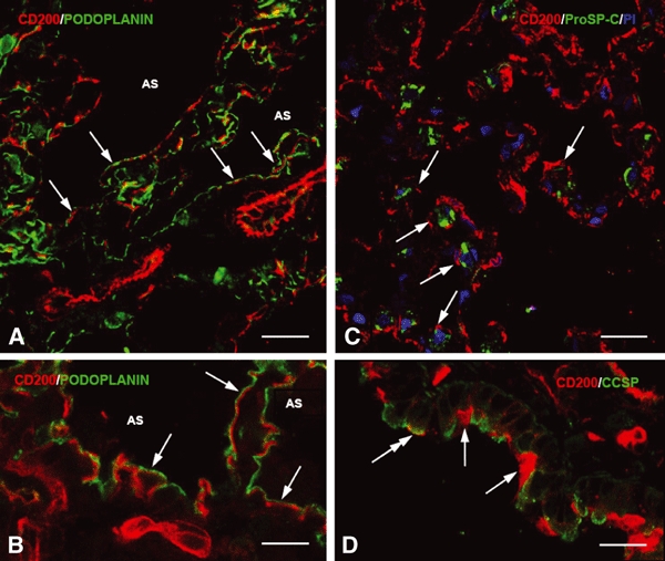
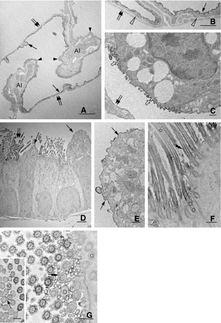
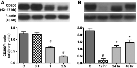
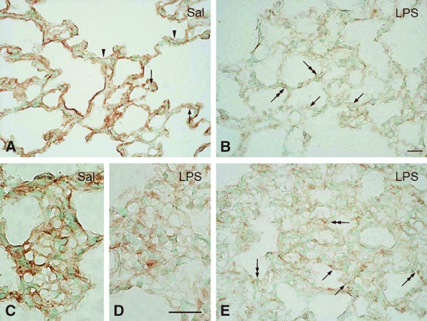
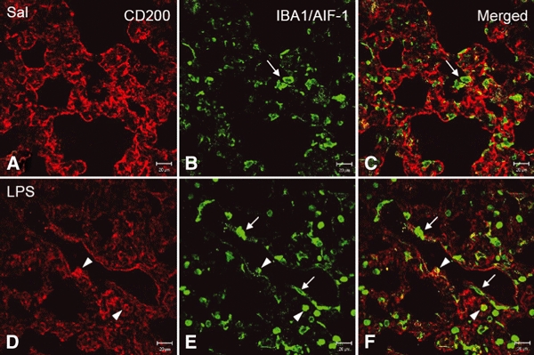
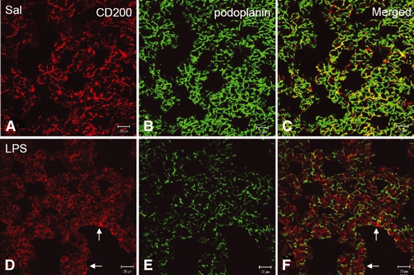
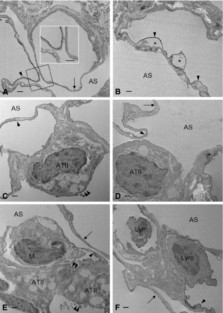
In vivo and in vitro studies have clearly demonstrated that signaling mediated by the interaction of CD200 and its cognate receptor, CD200R, results in an attenuation of inflammatory or autoimmune responses through multiple mechanisms. The present results have shown a differential expression of CD200 in the respiratory tract of intact rats. Along the respiratory passage, CD200 was specifically distributed at the bronchiolar epithelia with intense CD200 immunoreactivity localized at the apical surface of some ciliated epithelial cells; only a limited expression was detected on the Clara cells extending into the alveolar duct. In the alveolar septum, double immunofluorescence showed intense CD200 immunolabeling on the capillary endothelia. A moderate CD200 labeling was observed on the alveolar type II epithelial cells. It was, however, absent in the alveolar type I epithelial cells and the alveolar macrophages. Immunoelectron microscopic study has revealed a specific distribution of CD200 on the luminal front of the thin portion of alveolar endothelia. During endotoxemia, the injured lungs showed a dose- and time-dependent decline of CD200 expression accompanied by a vigorous infiltration of immune cells, some of them expressing ionized calcium binding adapter protein 1 or CD200. Ultrastructural examination further showed that the marked reduction of CD200 expression was mainly attributable to the loss of alveolar endothelial CD200. It is therefore suggested that CD200 expressed by different lung cells may play diverse roles in immune homeostasis of normal lung, in particular, the molecules on alveolar endothelia that may control regular recruitment of immune cells via CD200-CD200R interaction. Additionally, it may contribute to intense infiltration of immune cells following the loss or inefficiency of CD200 under pathological conditions.


Figures

Fig. 1
Fig. 1
(A–E) CD200 immunoreactivity in the conducting airways of intact rat lungs. CD200 labeling is localized consistently at the blood vessels (A–B, BV) along the airways but is absent at the epithelia of the nasal cavity, trachea (A, arrows) and bronchus (B, arrows). CD200 immunoreactivity of varying intensity appears initially at the bronchioles, particularly at specific groups of the ciliated epithelial cells (C, inset, arrows) and Clara cells (D, inset, arrows); some cells are CD200-negative (C–D, inset, double-headed arrows). In the alveolar tissues, CD200 staining along the alveolar septum appears discontinuous (E, arrows). Other cells/structures are not clearly defined as CD200-positive except for some type II pneumocytes which show a unique vacuolated cytoplasm containing weak but detectable CD200 immunoreactive product (E, double-headed arrow). Other epithelial cells (E, arrowheads) and alveolar macrophages (E, open arrowheads) are clearly negative for CD200. Scale bars: 20 μm.
Fig. 2
Fig. 2
(A–D) Immunofluorescence micrographs showing double labeling of CD200 (red) with podoplanin (alveolar type I cell marker, A–B, green), pro-surfactant-C (alveolar type II cell marker, C, green) or Clara cell secretory protein (Clara cell maker, D, green). Note that most CD200 immunoreactivity does not overlap with podoplanin, which appears to lie parallel to or above CD200 (A–B, arrows) and faces the airspace (AS). After staining with the nuclear dye propidium iodide (PI), alveolar type II cells show granule-like immunoreactivity of pro-surfactant-C and are incompletely outlined by weak CD200 immunoreactivity (C, arrows). In D, most Clara cells exhibit very weak CD200 labeling amongst the intensely-labeled cilia (arrows). Only a Clara cell shows significant CD200 on its apical membrane (D, double-headed arrow). Scale bars: (A,C–D) 20 μm, (B) 10 μm.
Fig. 3
Fig. 3
(A–G) Electron micrographs showing CD200 immunoreactivity in intact rat lungs. A preferential distribution of CD200 is observed at the luminal cytoplasmic membrane (A,B, arrows) of alveolar endothelia forming blood–air barrier. CD200 is negligible or absent on the remaining front of endothelia (A, arrowheads) or on the area that is facing the alveolar interstitium (A, AI). Moreover, the abluminal wall of alveolar endothelia (B, open arrowheads) and the alveolar type I epithelia (A–C, double arrows) were totally negative for CD200. Alveolar type II epithelial cells show CD200 expression exclusively on their apical cytoplasmic membrane furnished with short microvilli (C,). In the conducting airways, CD200 reaction product is distributed at the apical membrane of Clara cells (D–E, arrows) and cilia (D, double arrows) on the bronchiolar epithelia. Some cilia show a progressively increased CD200 immunoreactivity along the microtubule bundle in an apical to basal gradient (F). Microtubule bundles or axonemes constituting the most apical part (F, inset, ◊), base (F–G, □) and connecting basal body (F–G, ○) of the cilium show lack of CD200. The ciliary membrane also exhibits CD200 that is confined to the most apical (inset in G, arrow) and basal (F–G, double-headed arrows) parts; it is, however, inconsistent at the middle region of the cilium. Scale bars: (D) 2 μm; (E–G), inset in (G) 0.5 μm.
Fig. 4
Fig. 4
(A–B) Western blot analysis of CD200 expression in rat lung. Adult rats are intratracheally instilled with an increasing concentration (0.1, 1 or 2.5 mg kg−1) of LPS for 24 h (A), or others with 1 mg kg−1 LPS for different time intervals (12, 24 or 48 h) (B). Representative Western blots are shown in upper frames and relative levels of CD200 expression determined by densitometric scanning of the CD200 bands and normalized to the β-actin signal are shown in the bar graphs. Data shown are the mean ± SE of at least three separate experiments and are presented as the CD200/β-actin ratio. Statistical significance is established by anova followed by post hoc Tukey's multiple comparison tests. #P < 0.01 vs. each group; *P < 0.01 vs. the control or 12 h-treatment of LPS. In rats treated with different doses of LPS (A), lung CD200 is significantly reduced after treatment with 1–2.5 mg kg−1 LPS, whereas 0.1 mg kg−1 does not change the expression. The significant reduction of lung CD200 is evident as early as 12 h post-challenge with 1 mg kg−1 LPS (B). The CD200 reduction continues and expression is restored at 24 and 48 h post-challenge, but the expression levels at these time intervals are not statistically different (B).
Fig. 5
Fig. 5
(A–E) CD200 expression in the alveolar sac of rats intratracheally instilled with saline (Sal) or LPS for 24 h. In the Sal-treated (A) lung, CD200 immunoreactive product is clear but discontinuous along the alveolar septum (A, arrowheads). Weak immunoreactivity is found on alveolar type II epithelial cells with vacuolated cytoplasm (A, arrows). After LPS challenge (B), CD200 immunoreaction product is markedly reduced and appears attenuated along the septum where ATII shows a similar and weak immunoreactivity (B, arrows). Numerous lymphocyte-like cells (B, double-headed arrows) with a dense nucleus are clearly positive for CD200 and occupy the septum. In a tangential section of the septum, an obvious capillary network with intense immunoreactivity is observed in the Sal-treated lung (C); this becomes inconspicuous after LPS instillation (D). In areas with severe injury, the widened alveolar septum is infiltrated with more immune cells, including some cells which are CD200-positive (E, double-headed arrows). ATII expressing CD200 become more obvious in the pale background of injured tissues with reduced CD200 (E, arrows). Scale bars: 50 μm.
Fig. 6
Fig. 6
(A–F) Expression of IBA1/AIF-1 and CD200 in lungs of rats treated with intratracheally instilled saline or LPS for 12 h. In the control or saline (A–C, Sal)-treated lungs, IBA1/AIF-1 marks immune cells including macrophages and T lymphocytes (B–C, arrows) in either the alveolar septum or the airspace. The labeled cells are irregular in outline. Following LPS challenge, CD200 immunoreactivity is reduced (D) and more IBA1/AIF-1-labeled cells are found, especially those which are long and irregular around the blood vessel (E–F, arrows), and round cells in the thickened septum and the lumen of blood vessel. Some IBA1/AIF-1-positive round cells unexpectedly express CD200 (E,F, arrowheads). Scale bars: 20 μm.
Fig. 7
Fig. 7
(A–F) Expression of podoplanin and CD200 in lungs of rats intratracheally instilled with saline or LPS for 12 h. In the saline-treated (A–C, Sal) rats, patchy but intense CD200 immunoreactivity (A) is associated with podoplanin reaction products (B) in the control alveolar tissues. After LPS challenge, CD200 immunoreactivity diminishes and appears as diffused pattern with some positive cells in the thickened alveolar septum (D,F, arrows). Note that podoplanin immunoreactivity (B–C) is also noticeably reduced after LPS challenge (E,F). Scale bars: 20 μm.
Fig. 8
Fig. 8
(A–F) Electron micrographs showing CD200 expression in the distal airway of rat lungs challenged with LPS. CD200 is normally expressed on the luminal membrane of the alveolar endothelial cells, specifically on the thin portion that constitutes the blood–air barrier. A similar expression of CD200 is evidenced on the endothelia challenged with LPS (A–F, arrowheads). LPS markedly down-regulates CD200 expression on alveolar endothelia, which show lack of CD200 (A,D–F, arrows) or signs of degeneration with vacuolated cytoplasm (B, *). Endotoxin challenge does not affect CD200 expression (E, double arrowheads) significantly on alveolar type II epithelial cells (ATII); some of them display reduced amounts of CD200 (C, double arrowheads), whereas other display a total lack of CD200 (D). A monocytic cell (E,M), negative for CD200, is seen closely adherent to the thin portion of affected alveolar endothelium. The close cell contact is also evident between lymphocytes (F, Lym) and endothelial cells, both of which are CD200-positive. (Insert in A) Higher magnification of the structure outlined. AS, airspace. Scale bars: 500 nm.

Similar articles

Cited by

References

    1. Alam T, An MR, Papaconstantinou J. Differential expression of three C/EBP isoforms in multiple tissues during the acute phase response. J Biol Chem. 1992;267:5021–5024. - PubMed
    1. Aoki T, Matsumoto Y, Hirata K, et al. Expression profiling of genes related to asthma exacerbations. Clin Exp Allergy. 2009;39:213–221. - PubMed
    1. Arndt U, Wennemuth G, Barth P, et al. Release of macrophage migration inhibitory factor and CXCL8/interleukin-8 from lung epithelial cells rendered necrotic by influenza A virus infection. J Virol. 2002;76:9298–9306. - PMC - PubMed
    1. Barclay AN, Clark MJ, McCaughan GW. Neuronal/lymphoid membrane glycoprotein MRC OX-2 is a member of the immunoglobulin superfamily with a light-chain-like structure. Biochem Soc Symp. 1986;51:149–157. - PubMed
    1. Bartling B, Hofmann HS, Weigle B, et al. Down-regulation of the receptor for advanced glycation end-products (RAGE) supports non-small cell lung carcinoma. Carcinogenesis. 2005;26:293–301. - PubMed

Publication types

LinkOut - more resources