Maternal and fetal microvasculature in sheep placenta at several stages of gestation
- PMID: 20070427
- PMCID: PMC2829387
- DOI: 10.1111/j.1469-7580.2009.01184.x
Maternal and fetal microvasculature in sheep placenta at several stages of gestation
Abstract
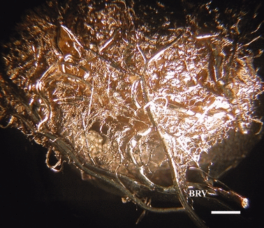
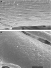
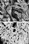
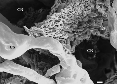
Maternal and fetal microvasculature was studied in ewes at days 50, 90 and 130 of gestation using microvascular corrosion casting and scanning electron microscopy. Microvascular corrosion casts of caruncles at day 50 were cup-shaped with a centrally located cavity. Branches of radial arteries entered the caruncle from its base and ramified on the maternal surface of the caruncle. Stem arteries broke into an extensive mesh of capillaries forming crypts on the fetal surface. The architecture of the caruncle at day 90 was similar to what was found at day 50 but the vascularity and the depth of the crypts increased in correspondence to increased branching of fetal villi. The substance of the caruncle was thicker at day 130 compared with day 50, with no remarkable difference compared with day 90. Capillary sinusoids of irregular form and diameter were observed on the fetal surface of the caruncle at all stages. These sinusoids may reduce blood flow resistance and subsequently increase transplacental exchange capacity. A microvascular corrosion cast of the cotyledon was cup-shaped with wide and narrow sides. Cotyledonary vessels entered and left the cotyledon from the narrow side. A cotyledonary artery gave proximal collateral branches immediately after entering the cotyledon and then further branched to supply the remaining portion of the cotyledon. Vessel branches broke into a mesh of capillaries forming the fetal vascular villi. Fetal villi that were nearest to the center of the cotyledon were the longest. Capillaries forming villi were in the form of a web-like mesh, were irregular in size and had sinusoidal dilations. The architecture of the cotyledon at day 90 was similar to day 50, but the vascularity increased. Branching of the fetal villi became more abundant. This extensive branching presumably allows a higher degree of invasion and surface contact to maternal tissues. At day 130, the distal portions of the fetal villi showed low ridges and troughs to increase the surface area for diffusion. Branching of fetal villi appears to influence the elaboration of maternal crypts in all stages of gestation. However, correspondence between crypts and villi is restricted to distal portions of fetal villi.
Figures


References
-
- Barcroft J, Barron DH. Observation upon the form and relations of the maternal and fetal vessels in the placenta of the sheep. Anat Rec. 1946;94:569–595. - PubMed
-
- Barker DJ, Clark PM. Fetal undernutrition and disease in later life. Rev Reprod. 1997;2:105–112. - PubMed
-
- Borowicz PP, Arnold DR, Grazul-Bilaska AT, et al. Modeling vascular growth in the sheep placentome. Biol Reprod. 2003;68(Suppl. 1):150.
-
- Borowicz PP, Arnold DR, Johnson ML, et al. Placental growth throughout the last two-thirds of pregnancy in sheep: vascular development and angiogenic factor expression. Biol Reprod. 2007;76:259–267. - PubMed
-
- Borowicz PP, Hafez SA, Redmer DA, et al. Chapter 10. Methods for evaluating uteroplacental angiogenesis and their application using animal models. In: Cheresh D, editor. Methods in Enzymology, Vol. 445, Angiogenesis, In Vivo, Part B. NY: Elsevier; 2008. pp. 229–253. - PubMed
Publication types
MeSH terms
Grants and funding
LinkOut - more resources
Full Text Sources
Miscellaneous

