

Magnetic resonance imaging performed on a hydrated mummy of medieval Korea
- PMID: 20070429
- PMCID: PMC2829391
- DOI: 10.1111/j.1469-7580.2009.01185.x
Magnetic resonance imaging performed on a hydrated mummy of medieval Korea
Abstract
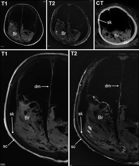
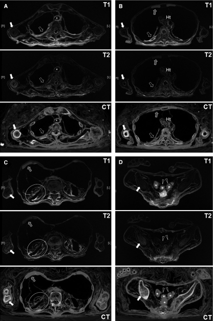
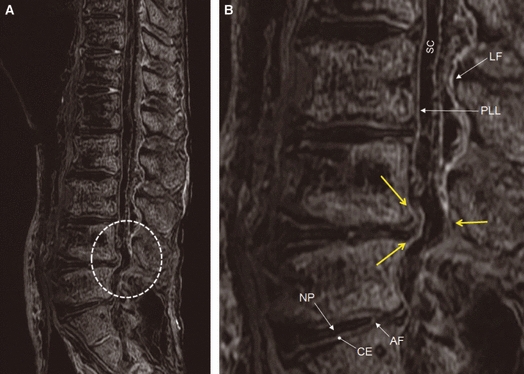
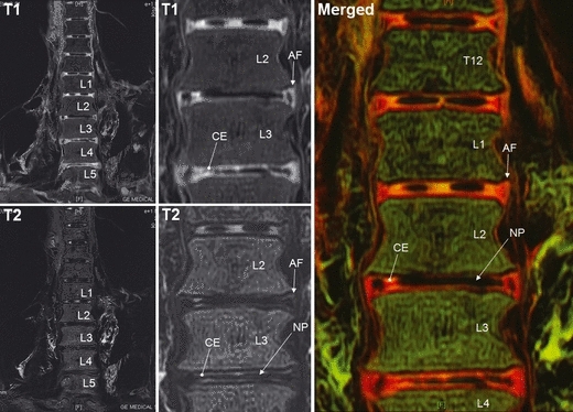
Previous investigations have shown that magnetic resonance imaging (MRI) can be employed as an efficient non-invasive diagnostic tool in studies on Egyptian mummies. MRI, moreover, because it produces especially clear images of well-hydrated tissue, could be a particularly effective diagnostic option for mummies that still retain humidity within tissues or organs. Therefore, in the present study, we tested MRI on a 17th century mummy, one of the most perfectly preserved 'hydrated mummies' ever found in Korea, in order to determine the quality of images that could be obtained. We found that the diagnostic value of an MRI scan of the hydrated mummy was not inferior to that of a computed tomography scan. The T1- and T2-weighted magnetic resonance (MR) signals showed unique patterns not easily obtained by computed tomography, the resultant MR images revealing the organ specificities clearly. Overall, the quality of the MR images from the hydrated mummy was superb and the scientific value of MRI in the study of hydrated mummies should not be underestimated.
Figures
Similar articles
-
Magnetic resonance imaging for the study of mummies.Magn Reson Imaging. 2016 Jul;34(6):785-794. doi: 10.1016/j.mri.2016.03.012. Epub 2016 Mar 12. Magn Reson Imaging. 2016. PMID: 26979539 Review.
-
Radiological analysis on a mummy from a medieval tomb in Korea.Ann Anat. 2003 Jul;185(4):377-82. doi: 10.1016/S0940-9602(03)80065-1. Ann Anat. 2003. PMID: 12924477
-
The potential for non-invasive study of mummies: validation of the use of computerized tomography by post factum dissection and histological examination of a 17th century female Korean mummy.J Anat. 2008 Oct;213(4):482-95. doi: 10.1111/j.1469-7580.2008.00955.x. J Anat. 2008. PMID: 19014355 Free PMC article.
-
A probable case of Hand-Schueller-Christian's disease in an Egyptian mummy revealed by CT and MR investigation of a dry mummy.Coll Antropol. 2012 Mar;36(1):281-6. Coll Antropol. 2012. PMID: 22816232
-
Mummies.Am J Phys Anthropol. 2007;Suppl 45:162-90. doi: 10.1002/ajpa.20728. Am J Phys Anthropol. 2007. PMID: 18046750 Review.
Cited by
-
Invasive versus Non Invasive Methods Applied to Mummy Research: Will This Controversy Ever Be Solved?Biomed Res Int. 2015;2015:192829. doi: 10.1155/2015/192829. Epub 2015 Aug 6. Biomed Res Int. 2015. PMID: 26345295 Free PMC article. Review.
-
Mummification in Korea and China: Mawangdui, Song, Ming and Joseon Dynasty Mummies.Biomed Res Int. 2018 Sep 13;2018:6215025. doi: 10.1155/2018/6215025. eCollection 2018. Biomed Res Int. 2018. PMID: 30302339 Free PMC article. Review.
-
Modular Coils with Low Hydrogen Content Especially for MRI of Dry Solids.PLoS One. 2015 Oct 23;10(10):e0139763. doi: 10.1371/journal.pone.0139763. eCollection 2015. PLoS One. 2015. PMID: 26496192 Free PMC article.
-
Anatomical confirmation of computed tomography-based diagnosis of the atherosclerosis discovered in 17th century Korean mummy.PLoS One. 2015 Mar 27;10(3):e0119474. doi: 10.1371/journal.pone.0119474. eCollection 2015. PLoS One. 2015. PMID: 25816014 Free PMC article.
-
Radiological diagnosis of congenital diaphragmatic hernia in 17th century Korean mummy.PLoS One. 2014 Jul 2;9(7):e99779. doi: 10.1371/journal.pone.0099779. eCollection 2014. PLoS One. 2014. PMID: 24988465 Free PMC article.
References
-
- Andreasen C, Gullov HC, Hart-Hansen JP, et al. The Greenland Mummies. London: The Trustees of the British Museum; 1991.
-
- Fleckinger A. Ötzi, the Iceman. The Full Facts at a Glance. Vienna/Bolzano: Folio, and the South Tyrol Museum of Archaeology; 2007.
-
- Hess MW, Klima G, Pfaller K, et al. Histological investigations on the Tyrolean Ice Man. Am J Phys Anthropol. 1998;106:521–532. - PubMed
-
- Karlik SJ, Bartha R, Kennedy K, et al. MRI and multinuclear MR spectroscopy of 3,200-year-old Egyptian mummy brain. AJR Am J Roentgenol. 2007;189:105–110. - PubMed
Publication types
MeSH terms
LinkOut - more resources
Full Text Sources
Medical
